|

Linear endoscopic ultrasound (longitudinal endosonography) allows the
visualization of accessories in the ultrasonic field, thus permitting
the real-time control of EUS-guided fine needle aspiration (EUS-FNA) biopsy and other
interventional procedures. Recent EUS-directed or EUS-assisted techniques are continuously expanding the spectrum of EUS applications. Most of the interventional procedures are further shown in the linear EUS atlas.
The EUS examinations were performed in the Gastroenterology Department of the University of Medicine and Pharmacy Craiova, ROMANIA, using a high resolution digital linear endoscopic ultrasound system, as well as DVD recording and archiving of cases.
New EUS cases are added, courtesy of Prof. Dr. Peter Vilmann, Chief of Gastrointestinal Endoscopy Laboratory, Department of Gastrointestinal Surgery, Gentofte University Hospital and University of Copenhagen, DENMARK. 
|
|
|
February 13, 2026 |
Now including
222 cases |
You are visitor #
44850 |
Online users :
1 |
|
|
| Neuroendocrine tumor |
Feb 26, 2009 |
|
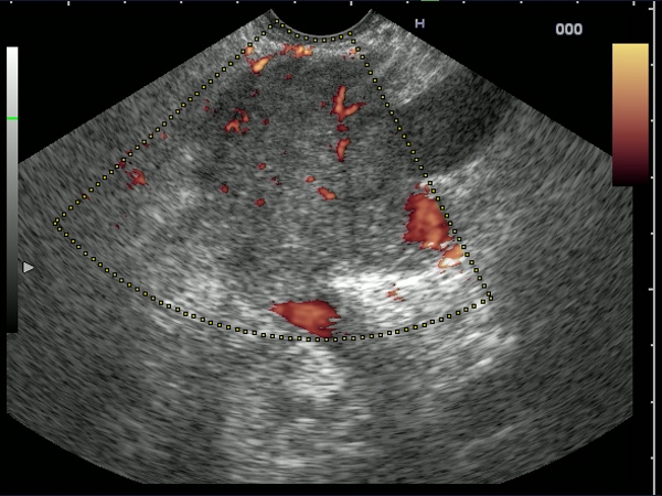
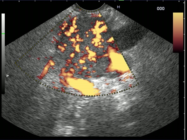
| Hypervascular tumor mass of the pancreatic tail visualized before and after contrast-enhancement (Sonovue 2.4 mL). The mass had predominant arterial signals inside, with a high pulsatility index. The diagnosis was confirmed by EUS-guided FNA with immunohistochemistry of the cell blocks. |
 |
 |
| |
More images / movies:


|
|
|
|
Contrast EUS > Pancreatic tumors > Malignant tumors |
|
| Pancreatic metastasis of malignant ocular melanoma |
Feb 26, 2009 |
|
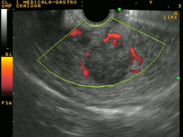
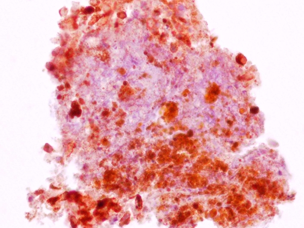
| A 43-years-old woman, diagnosed 4 years ago with a malignant ocular melanoma, removed by surgery and further treated by combined chemo-radiotherapy. Transabdominal ultrasound and contrast-enhanced CT showed a 20 mm tumor in the neck of the pancreas. EUS showed peripheral signals in power Doppler mode (Fig. 1). EUS-FNA with immunocytochemistry showed an intense cytoplasmatic and nuclear immunostaining for S100 and an intense cytoplasmatic immunostaining for HMB45 (Fig. 2). |
 |
 |
| |
More images / movies:


|
|
|
|
Pancreatic diseases > Pancreatic tumors > Metastatic tumors |
|
|
| |
|
| Dr. Adrian Saftoiu |
Gastroenterology Department |
Site concept, Case histories, Web layout |
| Dr. Adrian Saftoiu, Dr. Sergiu Cazacu |
Gastroenterology Department |
EUS examinations (images and movies) |
| Dr. Peter Vilmann |
Gentofte University Hospital and
University of Copenhagen |
EUS examinations (images and movies) |
Prof. Dr. Adrian Saftoiu, Prof. Dr. Tudorel Ciurea,
Prof. Dr. Ion Rogoveanu |
Gastroenterology Department |
Transabdominal ultrasound |
| Dr. Daniela Dumitrescu, Dr. Mihai Popescu |
Radiology Department |
CT, MRCP examinations |
| Dr. Adrian Saftoiu |
Gastroenterology Department |
ERCP examinations |
| Dr. Aristida Georgescu, Dr. Ernestina Andrei |
Radiology Department |
Radiology support for ERCP examinations |
| Dr. Carmen Popescu |
Cytology Laboratory |
Cytological diagnosis and EUS-FNA analysis |
| Dr. Claudia Valentina Georgescu |
Pathology Department |
Pathological diagnosis |
Dr. Sevastita Iordache, Dr. Dan-Ionuţ
Gheonea, Drd. Maria-Monalisa Filip |
Gastroenterology Department |
Case histories |
| Dr. Anca Maloş |
IC Unit |
EUS examinations (anesthesiology) |
| Drd. Ana-Maria Ioncică |
Pulmonology Department |
Case histories |
| Daniela Burtea, Monica Molete, Mihaela Caliţa |
Gastroenterology Department |
Endoscopy nursing |
| Eng. Gabriel Popescu, Eng. Cristina Cerbulescu |
IT Centre - U.M.F. Craiova |
Webmasters |
| Eng. Alex Iordache |
Arpanet.ro |
Site development |
Pictures are best viewed using at least High (32 bit) color quality and 1024x768 pixels, with Netscape 6.0 or higher, and Internet Explorer 6.0 or higher. Movies are usually 30 seconds long, compressed with DivX 5.20 or above (
download
). |


