| Neuroendocrine tumor |
Feb 26, 2009 |
|
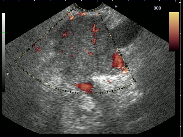
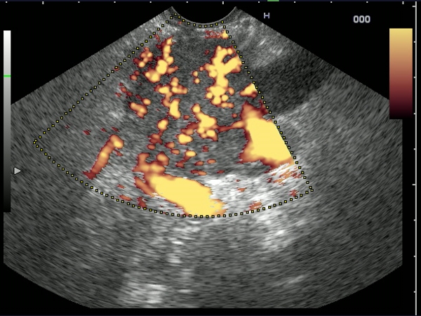
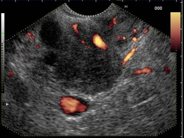
| Hypervascular tumor mass of the pancreatic tail visualized before and after contrast-enhancement (Sonovue 2.4 mL). The mass had predominant arterial signals inside, with a high pulsatility index. The diagnosis was confirmed by EUS-guided FNA with immunohistochemistry of the cell blocks. |
 |
 |
| |
More images / movies:


|
|
|
| 3D pancreatic adenocarcinoma |
Feb 22, 2009 |
|
| Tridimensional (3D) contrast-enhanced power Doppler EUS appearance of a malignant pancreatic body tumor, obtained 3 minutes after Sonovue injection, in the late phase. The splenomesenteric confluence and the portal vein can be easily visualized, as well as an increased collateral circulation. The diagnosis of pancreatic adenocarcinoma was confirmed by EUS-guided FNA. |
 |
 |
| |
|
|
|
| Pancreatic body adenocarcinoma |
Feb 22, 2009 |
|
| A 70-years-old woman with a pancreatic body adenocarcinoma, confirmed by EUS-guided FNA. The tumor had slight power Doppler signals before or after contrast-enhancement (Sonovue 2.4 mL), with enlarged collateral circulation around the tumor mass. |
 |
 |
| |
More images / movies:

|
|
|
| Advanced pancreatic body adenocarcinoma |
Feb 22, 2009 |
|
Large pancreatic body mass, in a 72-years-old man admitted with epigastric pain, weight loss and recent onset diabetes.
Vascular index is easily calculated during the late phase, after contrast-enhancement with Sonovue (2.4 mL), showing a relatively hypovascular tumor. The diagnosis of pancreatic adenocarcinoma was confirmed by EUS-guided FNA. |
 |
 |
| |
More images / movies:

|
|
|
| Pancreatic adenocarcinoma |
Feb 15, 2009 |
|
| Patient with a hypoechoic pancreatic head mass of approximately 40/30 mm, visualized by EUS in a 70-years-old women. The mass was invading the pancreaticoduodenal artery, having a low vascular index in power Doppler mode, both before, and also after contrast-enhancement (Sonovue 2.4 mL). |
 |
 |
| |
More images / movies:


|
|
|
|


