| Large ampullary carcinoma |
Feb 26, 2009 |
|
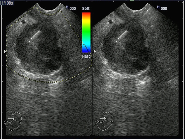
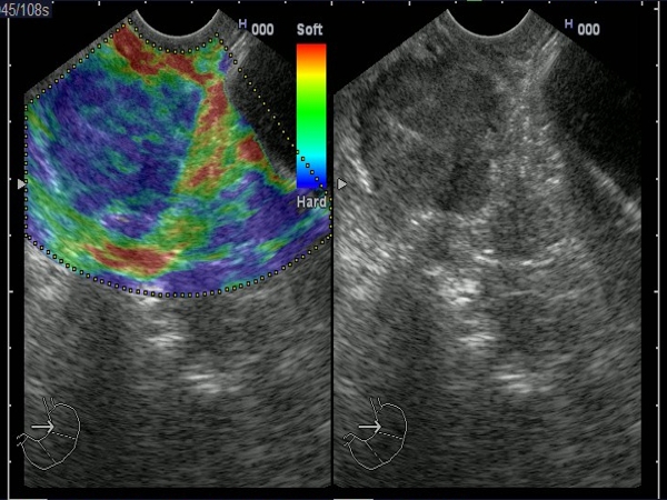
| A 64-years-old woman presented with intermittent jaundice and anemia. EUS showed a large mass projected in the papillary area, invading the pancreatic parenchyma. EUS elastography showed the mass as hard (blue). The diagnosis was confirmed by EUS-guided FNA, which showed an adenocarcinoma. |
 |
 |
| |
|
|
|
| Ampullary carcinoma |
Feb 5, 2005 |
|
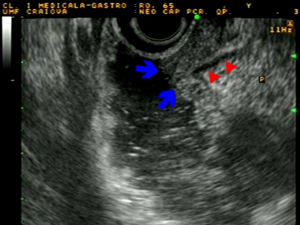
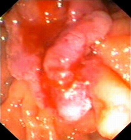
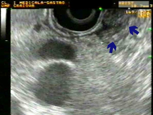
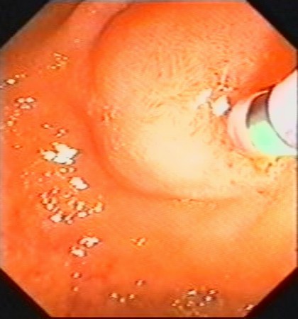
| Ulcerated tumor visualized as a hypoechoic mass at the level of major papilla (arrows), with integrity of the muscle layer (arrow heads) (T2). The tumor was identified with a lateral viewing duodenoscope as an ulcerated mass at the level of the ampula Vater; biopsies confirmed the diagnosis of moderately differentiated adenocarcinoma. |
 |
 |
| |
More images / movies:

|
|
|
| Intramural ampullary carcinoma |
Feb 5, 2005 |
|
| Linear EUS identified a hypoechoic tumor (16 mm) at the level of major papilla (arrows), with invasion of the pancreatic parenchyma (T3). The major papilla was subsequently visualized with a lateral viewing duodenoscope, having a normal covering mucosa. However, biopsies taken after biliary sphincterotomy confirmed the diagnosis of adenocarcinoma. |
 |
 |
| |
|
|
|
|


