| Gastric lymphoma |
Feb 18, 2009 |
|
| Patient with a B-cell lymphoma with recurrence after chemotherapy, with irregular and enlarged gastric folds, with multiple ulcerations. Endoscopic ultrasound (including miniprobes) showed large gastric folds, with a disorganized structure, with predominant involvement of the superficial and deep mucosa (EUS layers 1 and 2). Enlarged gastric wall (up to 10 mm), especially on the greater curvature, was visualized with miniprobes. |
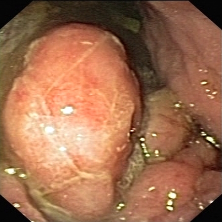 |
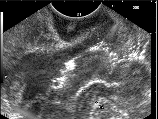 |
| |
More images / movies:


|
|
|
| Non-Hodgkin gastric lymphoma |
Sep 10, 2005 |
|
| Patient diagnosed with non-Hodkin primary gastric lymphoma, with an antral protruding mass masquerading a submucosal tumor assessed by EUS for staging. EUS showed a hypoechoic infiltration of EUS layers 2,3,4 with an enlarged, infiltrated gastric wall of up to 10 mm (T3 tumor). Multiple hypoechoic, homogenous, round-oval lymph nodes of up to 20 mm, were also visualized (N2). The patient was further reffered for complex chemotherapy. |
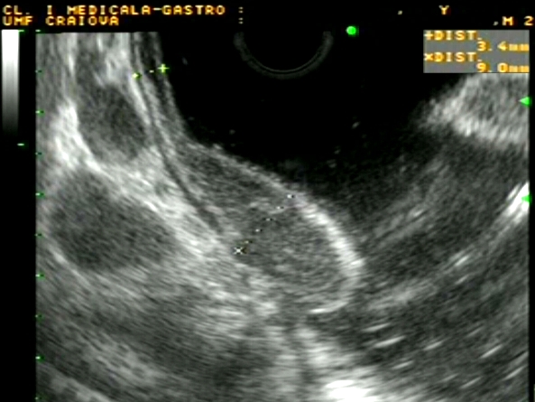 |
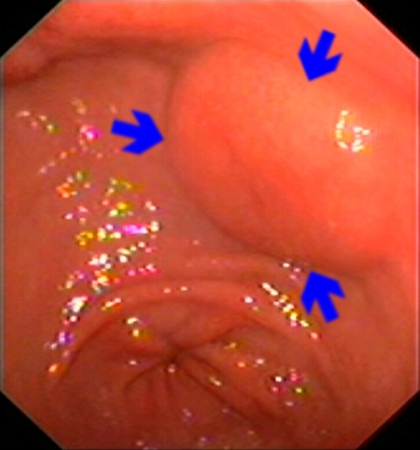 |
| |
More images / movies:


|
|
|
| Low-grade MALT gastric lymphoma |
Jan 23, 2005 |
|
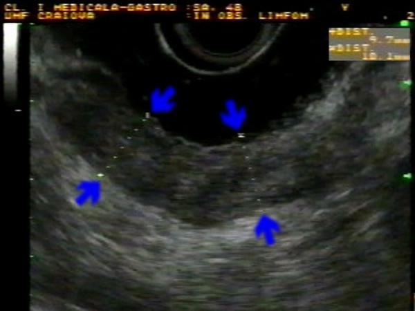
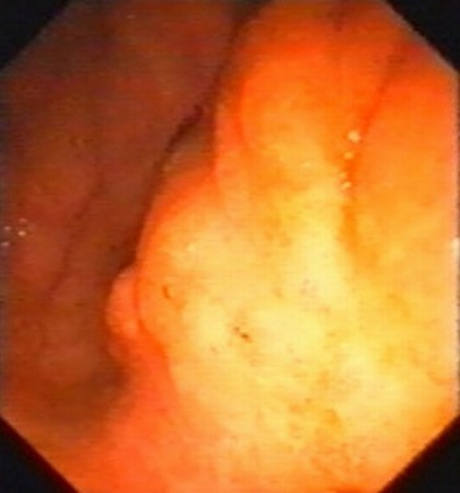
| Patient with biopsy proven gastric low-grade MALT lymphoma, with two episodes of upper gastrointestinal bleeding in the past six months, having a proeminent and infiltrated mucosa in the corporeal region of the stomach. Linear EUS showed transmural hypoechoic infiltration (blue arrows), with invasion of serosa (T3). The gastric wall was thickened to approximately 10 mm. |
 |
 |
| |
|
|
|
| High-grade MALT gastric lymphoma |
Jan 23, 2005 |
|
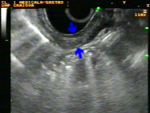
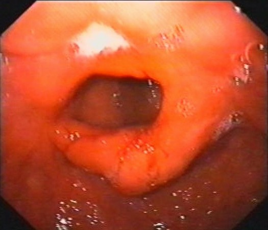
| Patient with biopsy proven gastric high-grade MALT lymphoma, with medio-gastric stenosis and multiple ulcerations. Linear EUS showed transmural hypoechoic infiltration (blue arrows), with invasion of serosa (T3). The gastric wall was thickened to approximately 8 mm. |
 |
 |
| |
|
|
|
| Low-grade MALT gastric lymphoma |
Jan 23, 2005 |
|
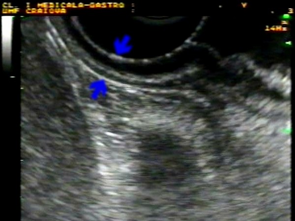
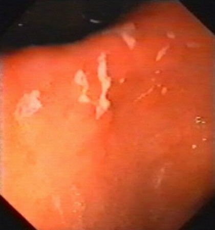
| Patient with biopsy proven gastric low-grade MALT lymphoma, with partial remission after H. pylori eradication treatment and chemotherapy. Multiple erosions were visualised by endoscopy in the antral region and on the gastric angle. Linear EUS showed a slightly thickened gastric wall (7 mm), with complete normalisation of the wall structure (5 layers). |
 |
 |
| |
|
|
|
|


