| Advanced lung cancer |
Feb 19, 2009 |
|
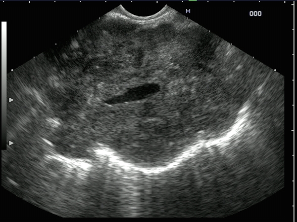
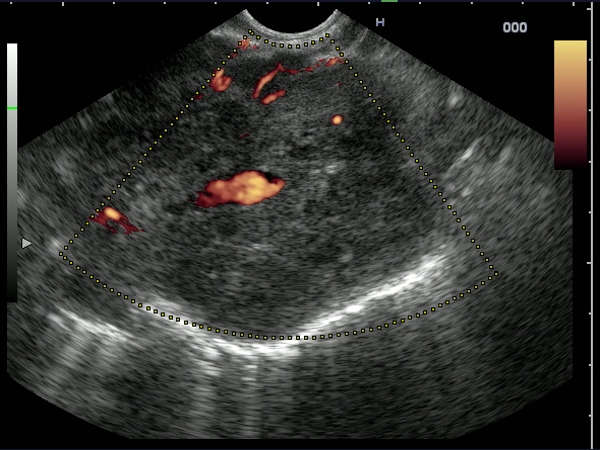
| Large hypoechoic, inhomogenous mediastinal mass, with peripheral hyperechoic echoes suggestive of air. The central anechoic region coresponds to a large vessel, completely embedded by the tumor. The diagnosis was established by EUS-guided FNA from the tumor mass. |
 |
 |
| |
More images / movies:

|
|
|
| |
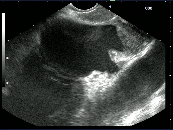
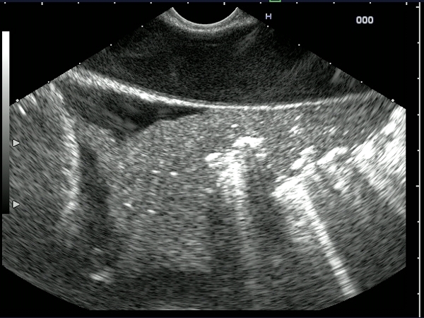
| Patient with a large mediastinal mass of 5 cm, confirmed by EUS-guided FNA as a squamous cell lung cancer. Multiple lymph nodes up to 3 cm were depicted in the aorto-pulmonary window. Invasion of the left atrium was clearly visible, as well as a pleural fluid collection (T4). |
 |
 |
| |
More images / movies:


|
|
|
| Lung cancer (T4) |
Feb 5, 2005 |
|
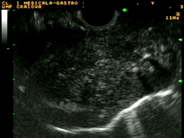
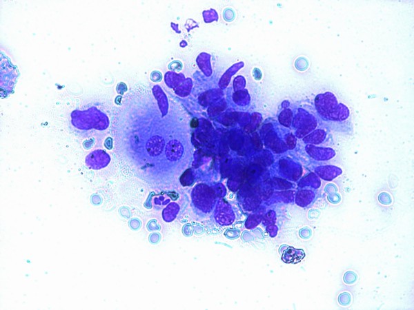
| Patient with biopsy-confirmed broncho-pulmonary carcinoma with mediastinal invasion visualized at 35 cm from the incisors. EUS-guided FNA established the diagnosis of malignancy, showing clumps of atypical cells. The patient was staged as T4NxMx (stage IIIb), being subsequently reffered to chemotherapy. |
 |
 |
| |
More images / movies:


|
|
|
|


