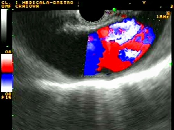
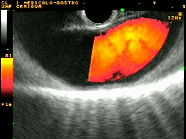
| Normal descending aorta |
Feb 11, 2005 |
|
| Patient with normal descending aorta visualized from the terminal esophagus in grey scale, color and power Doppler. The hyperechogenic echoes of the spine can be identified posteriorly. This is an important vascular structure, used for orientation and EUS examination of the mediastinum from the lower esophagus. |
 |
 |
| |
More images / movies:

|
|
|
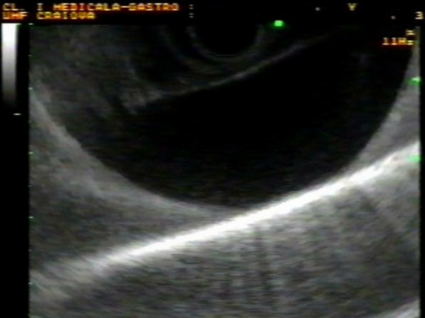
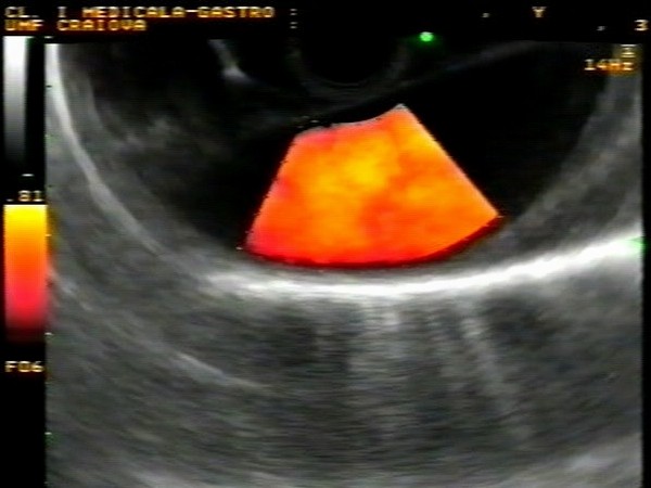
| Normal descending aorta |
Feb 9, 2005 |
|
| Normal descending aorta visualized from the terminal esophagus in grey scale and power Doppler. The hyperechogenic echoes of the spine can be easily identified posteriorly. This is an important vascular structure, used for orientation and EUS examination of the mediastinum from the lower esophagus. |
 |
 |
| |
|
|
|
|


