| Malignant neuroendocrine tumor |
Feb 23, 2009 |
|
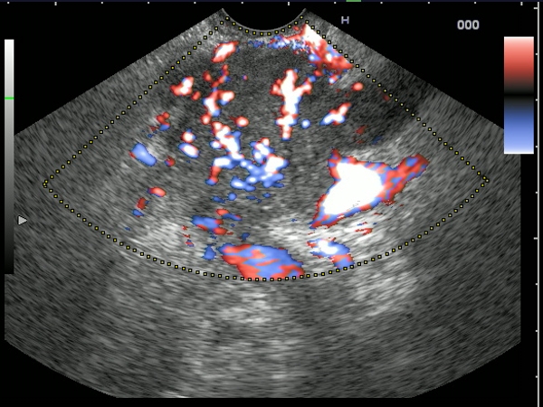
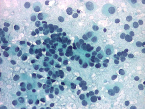
| A 61-years-old man with a hypoechoic, inhomogeneous mass of the pancreatic body, with rich pulsatile vascularization, especially visualized after contrast-enhanced EUS (Sonovue). The tumor invaded the celiac trunck, splenic artery and splenic vein (T4). Multiple peripancreatic lymph nodes were also visualized. EUS-guided FNA was performed and the cytological exam revealed small round cells with severe nuclear atypia. |
 |
 |
| |
More images / movies:


|
|
|
| Malignant neuroendocrine tumor |
Feb 23, 2009 |
|
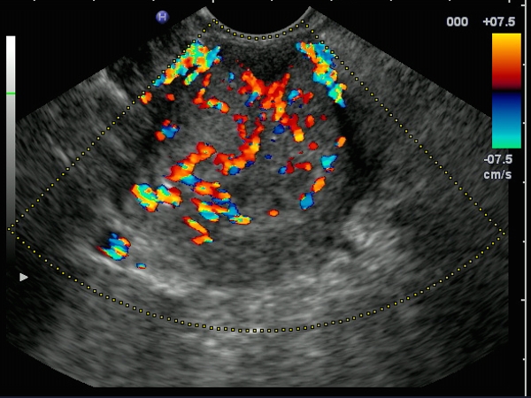
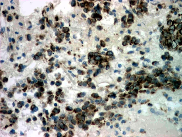
| Large tumor mass of the pancreatic tail discovered in a 55-years-old woman, visualized by EUS as a hypervascular mass in color Doppler or power Doppler mode. EUS-guided FNA established the diagnosis of neuroendocrine tumor, while immunohistochemistry of the cell-blocks was also positive. |
 |
 |
| |
More images / movies:
|
|
|
|


