| Serous cystadenoma |
Feb 10, 2009 |
|
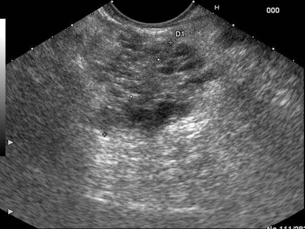
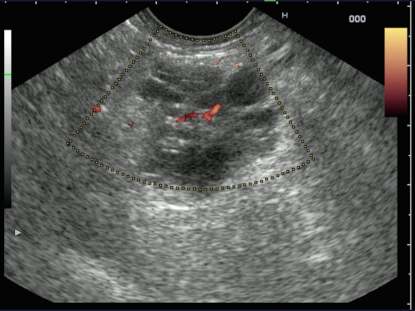
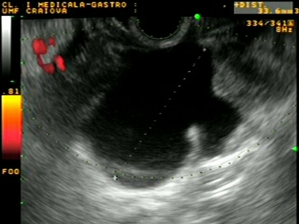
| A 71-years-old male patient, diagnosed by CT and abdominal ultrasonography with a pancreatic tumor mass. EUS revealed a 2.5 cm tumor, with multiple small cystic lesions inside, with discrete power Doppler signals visualized in the center. |
 |
 |
| |
More images / movies:

|
|
|
| Serous cystadenoma |
Feb 10, 2009 |
|
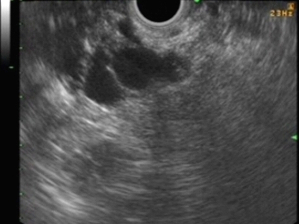
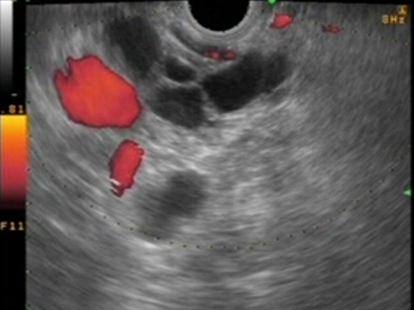
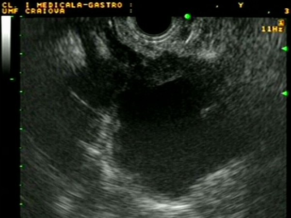
| A multicystic lesion in the pancreatic body / tail, of 2 centimeters diameter, visualized near the splenic vein. EUS-guided FNA and cytological exam revealed a benign etiology suggestive of a serous cystadenoma. |
 |
 |
| |
More images / movies:

|
|
|
| Pancreatic tail cystadenocarcinoma |
Aug 29, 2005 |
|
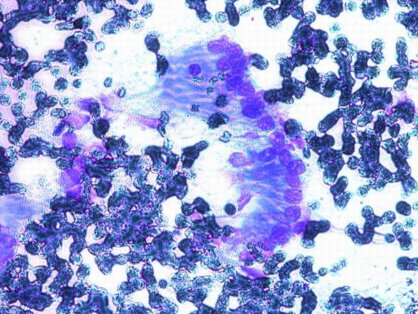
| Patient with a large cystic tumor mass of approximately 5 cm at the level of the pancreatic tail, with thickened and irregular walls. EUS-FNA with complete aspiration of the cystic fluid was initially performed, depicting very high values of CEA and CA 19-9. EUS-guided FNA of the solid part was subsequently done demonstrating cilindrical atypical cells with hyperchromatic nuclei and visible nucleoli. |
 |
 |
| |
More images / movies:
|
|
|
| Pancreatic tail cystadenocarcinoma |
Mar 2, 2005 |
|
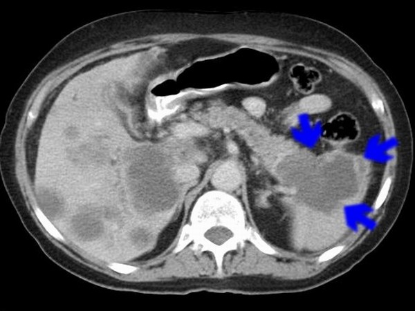
| Patient with a large cystic pancreatic tail tumor, visualized during CT. EUS of the pancreatic tail region showed the cystic tumor, with irregular and thick walls. EUS-FNA was performed through the stomach wall and cytological analysis was positive for atypical cells. Cystic fluid analysis also revealed high levels of CEA and CA 19-9. |
 |
 |
| |
More images / movies:
|
|
|
|


