| Severe chronic pancreatitis |
Dec 3, 2008 |
|
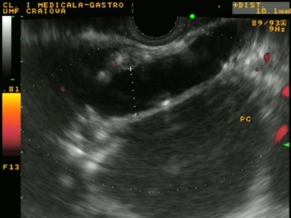
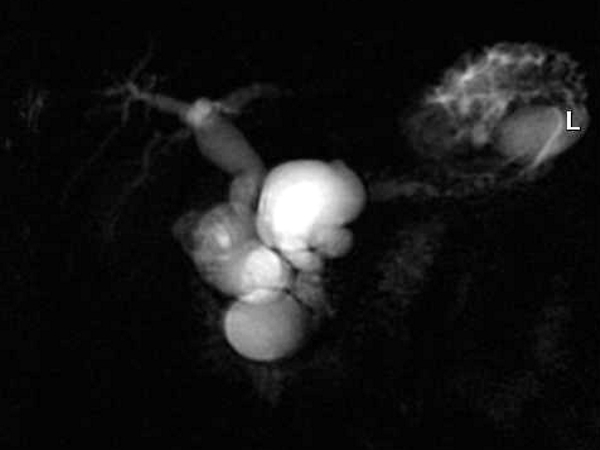
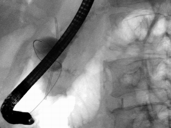
| A 60 year old male patient with chronic calcifying pancreatitis, with severe ductal obstruction and multiple communicating pancreatic pseudocysts. Dilated common bile duct, intrahepatic bile ducts and dilated main pancreatic duct (up to 10 mm) with multiple stones, as well as 3 pseudocysts at the level of the pancreatic head and one pseudocyst at the level of the pancreatic tail, were seen on both EUS and MRCP. |
 |
 |
| |
More images / movies:

|
|
|
| Pseudocyst drained by EUS |
Feb 9, 2005 |
|
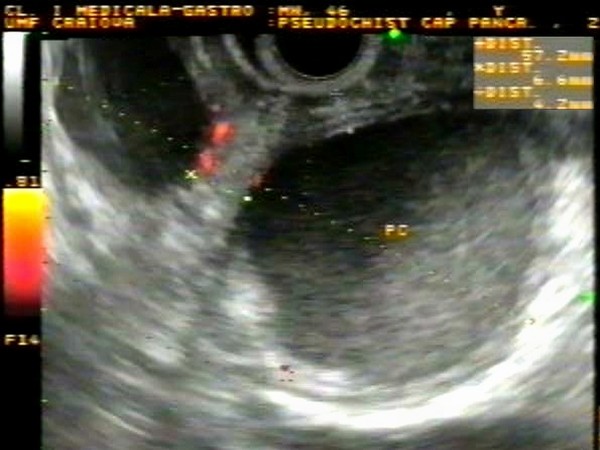
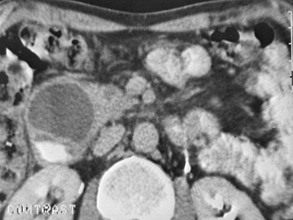
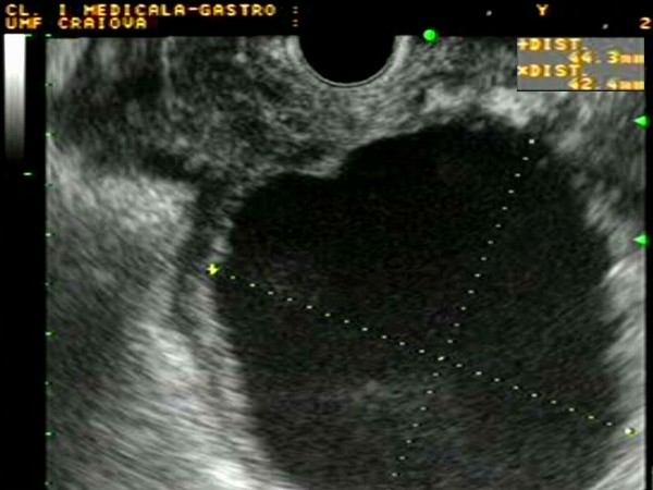
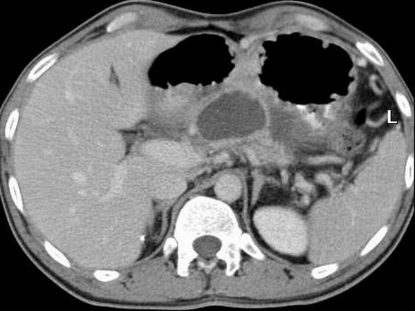
| Patient with a pancreatic head pseudocyst of approximately 6 cm, embedding and compressing the second duodenum. The distance between the duodenum and the pseudocyst content was less than 1 cm, safely allowing EUS-guided drainage. The pseudocyst can be seen on the corresponding CT image, with contrast inside the second duodenum. |
 |
 |
| |
More images / movies:

|
|
|
| Simple chronic pseudocyst |
Feb 5, 2005 |
|
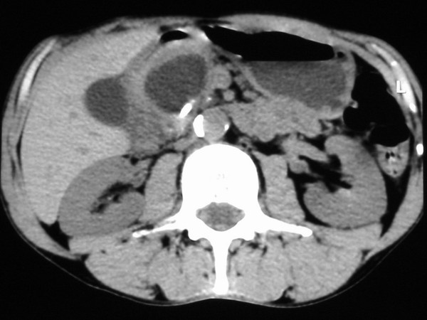
| Patient with chronic alcoholic pancreatitis with a pseudocyst of the body of the pancreas visualized on CT. EUS allowed the characterization of the content of the pseudocyst, the evaluation of the wall thickness, as well as the presence of the vascularization of the walls. |
 |
 |
| |
More images / movies:
|
|
|
| Pseudocyst with organized debris |
Feb 5, 2005 |
|
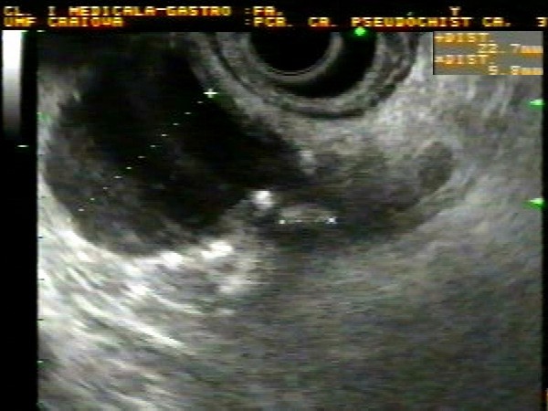
| Patient with a pancreatic pseudocyst with organized debris inside, disposed as small vesicles, visualized during linear EUS. The pseudocyst compressed the terminal common bile duct which was tortuos; both structures were visualized anterior to the the portal vein. The debris inside the pseudocyst was not visible on the corresponding CT image. |
 |
 |
| |
More images / movies:
|
|
|
| Pseudocyst drained by ERCP |
Jan 24, 2005 |
|
| Patient with a pancreatic head pseudocyst of approximately 2.5 cm, causing compression of the common bile duct and jaundice. EUS showed the pseudocyst with clear content and a small stone of 6 mm in the vicinity. ERCP showed direct communication between the pseudocyst and the dilated main pancreatic duct. Consequently, stents were placed both in the bile duct and the pseudocyst cavity, with resolution of the symptoms. |
 |
 |
| |
|
|
|
|


