| 3D advanced chronic pancreatitis |
Feb 26, 2009 |
|
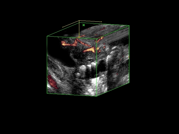
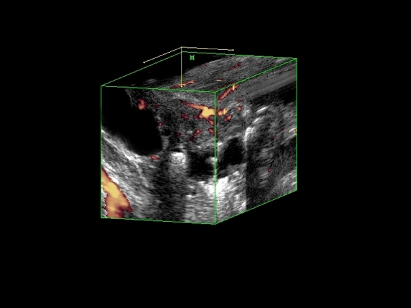
| Severe advanced chronic pancreatitis with double duct sign easily visible on 3D reconstructions: dilated common bile duct (20 mm), as well as a large pancreatic duct (8 mm) were easily visible, and caused by an impacted stone in the pancreatic duct. |
 |
 |
| |
|
|
|
| Advanced chronic pancreatitis |
Feb 24, 2009 |
|
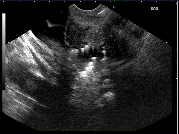
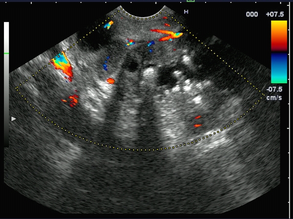
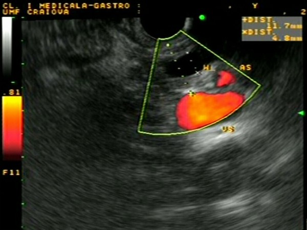
| Young patient (39 years-old), with long lasting chronic alcohol abuse, presenting for upper abdominal pain, jaundice and weight loss. EUS showed a severely altered pancreas, with intraductal and parenchymal calcifications. A dilated common bile duct (20 mm), as well as a large pancreatic duct (8 mm) were also visible. |
 |
 |
| |
More images / movies:

|
|
|
| Chronic obstructive pancreatitis |
Feb 5, 2005 |
|
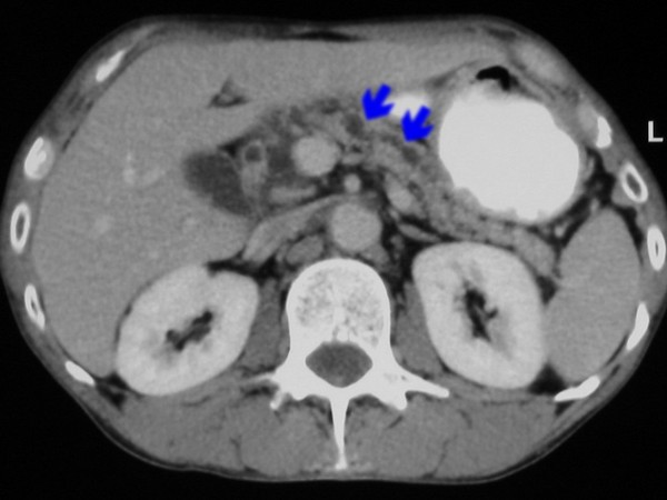
| Patient with chronic pancreatitis, with dilated main pancreatic duct at the level of the body of the pancreas, visualized during EUS from the gastric body, anterior to the splenic vessels (vein and artery). Dilated pancreatic duct was also visualized on the corresponding CT image. |
 |
 |
| |
More images / movies:
|
|
|
| Chronic calcifying pancreatitis |
Jan 24, 2005 |
|
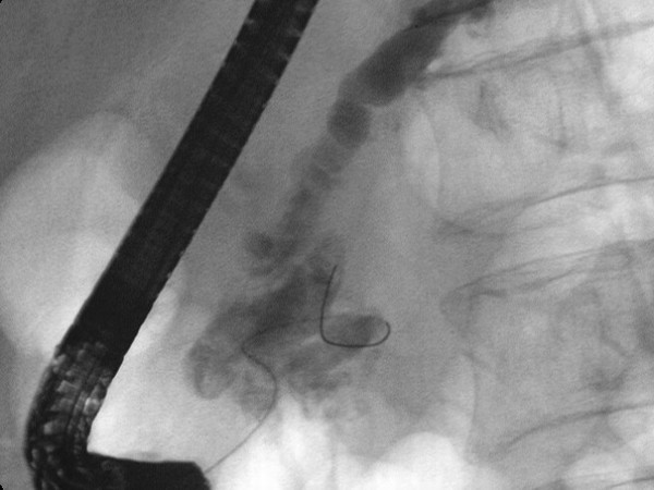
| Patient with heavy alcohol abuse, recently diagnosed with diabetes mellitus, examined by EUS. The pancreatic head was enlarged, inhomogeneous, with multiple stones with posterior shadow. ERCP showed the enlarged and disorganized pancreatic ducts, with multiple filling defects and calcifications in the pancreatic head. |
 |
 |
| |
|
|
|
|


