| Esophageal squamous cell carcinoma |
Feb 6, 2005 |
|
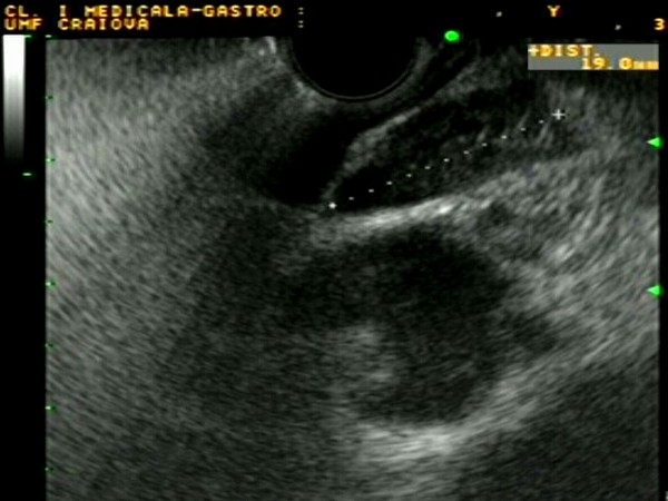
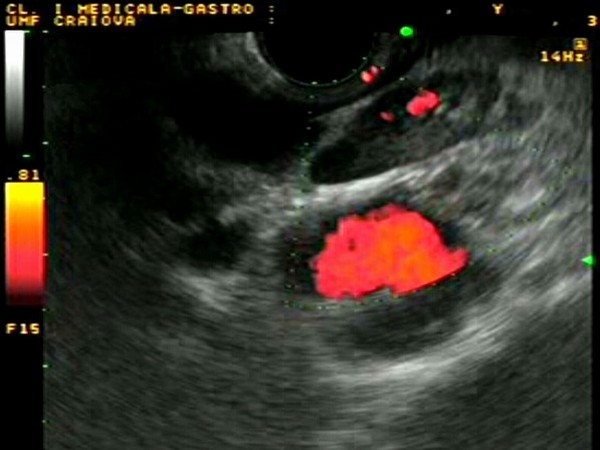
| Patient with esophageal squamous cell carcinoma located at 35 cm distance from the incisors. Linear EUS demonstrated enlarged mediastinal lymphnodes (up to 20 mm), located in the aorto-pulmonary window. The lymphnodes were oval, hypoechoic, homogenous, with peripheral and central vascular signals demonstrated in power Doppler mode (suggestive of a hilum). EUS-guided FNA confirmed that the lymphnodes were benign. |
 |
 |
| |
|
|
|
| Esophageal adenocarcinoma |
Feb 6, 2005 |
|
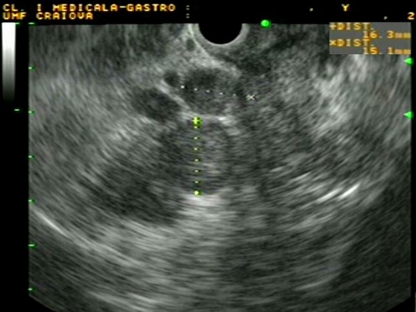
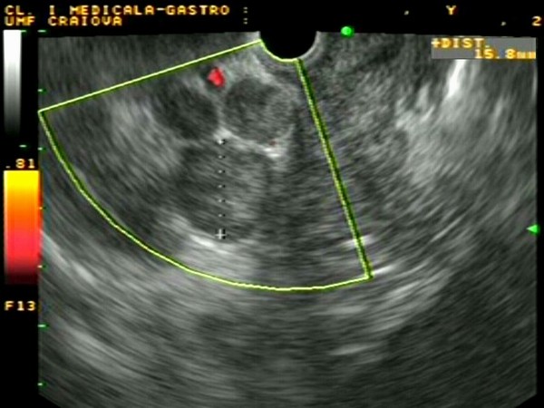
| Patient with adenocarcinoma located at 38 cm distance from the incisors. Linear EUS demonstrated enlarged mediastinal lymphnodes (up to 16 mm), located near the tumor and the celiac trunk. The lymhnodes were round, hypoechoic, inhomogenous, with peripheral halo and absence of vascular signals demonstrated in power Doppler mode. EUS-guided FNA confirmed that the lymphnodes were malignant. |
 |
 |
| |
|
|
|
|


