| Esophageal squamous cell carcinoma |
Jan 8, 2009 |
|
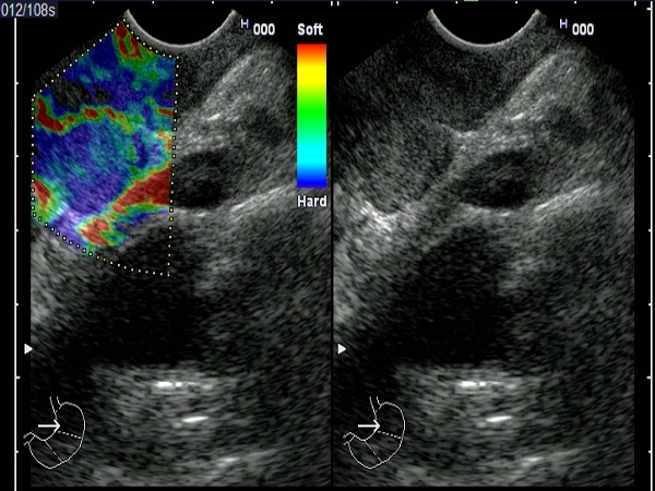
| Patient with a T4 esophageal squamous cell carcinoma at 30 cm from incisors, clearly invading the descending aorta. A peritumoral lymph node (16 mm) was depicted as hard by elastography. |
 |
 |
| |
More images / movies:

|
|
|
| Esophageal squamos cell carcinoma |
Feb 6, 2005 |
|
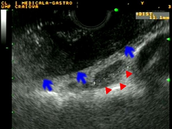
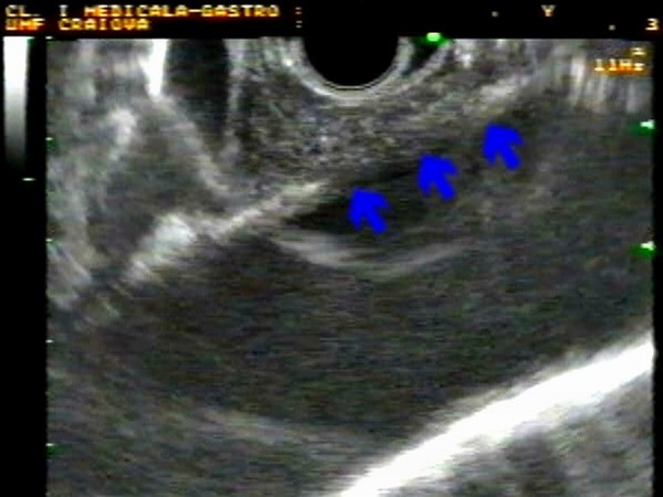
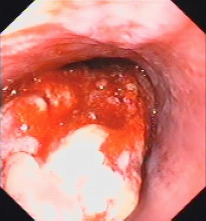
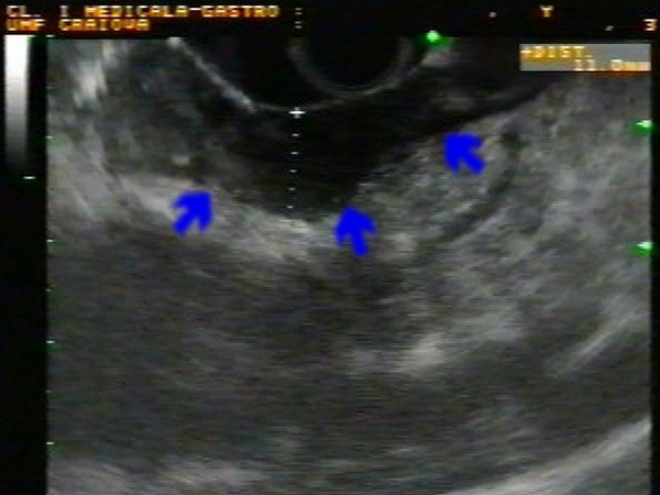
| Patient with esophageal squamous cell carcinoma located at 32 cm distance from the incisors. Linear EUS demonstrated an inhomogenous, hypoechogenic tumor (blue arrows), with disruption of the normal three-layer structure of the esophageal wall and invasion of the adventitia (T3). An enlarged oval lymphnode (red arrowheads) of 11 mm was seen adjacent to the tumor (N1). |
 |
 |
| |
|
|
|
| Esophageal adenocarcinoma |
Feb 6, 2005 |
|
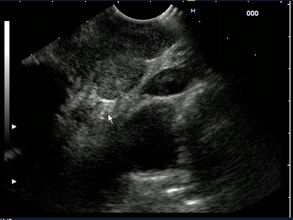
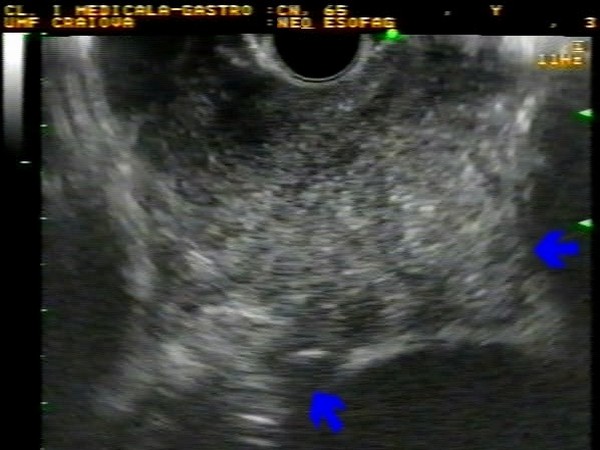
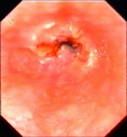
| Patient with esophageal adenocarcinoma located at 35 cm distance from the incisors. Linear EUS demonstrated an inhomogenous, hypoechogenic tumor, with disruption of the normal three-layer structure of the esophageal wall and invasion of the aorta (T4 carcinoma). |
 |
 |
| |
|
|
|
| Esophageal squamos cell carcinoma |
Feb 6, 2005 |
|
| Patient with stenosing esophageal squamos cell carcinoma located at 30 cm distance from the incisors. Linear EUS demonstrated an inhomogenous, hypoechogenic tumor, with disruption of the normal three-layer structure of the esophageal wall and invasion of the left atrium (T4 carcinoma). |
 |
 |
| |
|
|
|
| Esophageal squamous cell carcinoma |
Feb 6, 2005 |
|
| Patient with esophageal squamous cell carcinoma located at 30 cm distance from the incisors. Linear EUS demonstrated an inhomogenous, hypoechogenic tumor, with disruption of the normal three-layer structure of the esophageal wall and invasion of the adventitia (T3 carcinoma). |
 |
 |
| |
|
|
|


