| Malignant GIST |
Sep 9, 2005 |
|
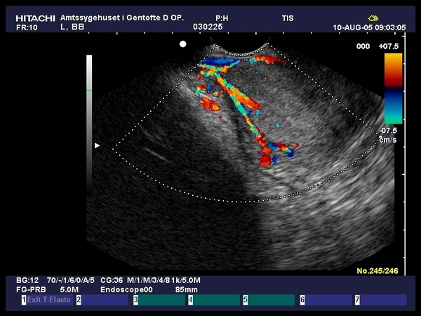
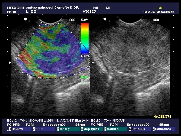
| Large gastric stromal tumor (GIST) identified by EUS as an 8 cm in-homogenous mass, with hypo- and hyper-echoic areas and imprecise margins. Color Doppler EUS depicted intense vascular signals in the periphery of the mass, predominantly of arterial origin. Elastography showed a hard in-homogenous tumor. Due to the strong suspicion of malignancy the patient was referred for surgery (provided by Prof. Dr. Peter Vilmann, Copenhagen). |
 |
 |
| |
More images / movies:


|
|
|
| |
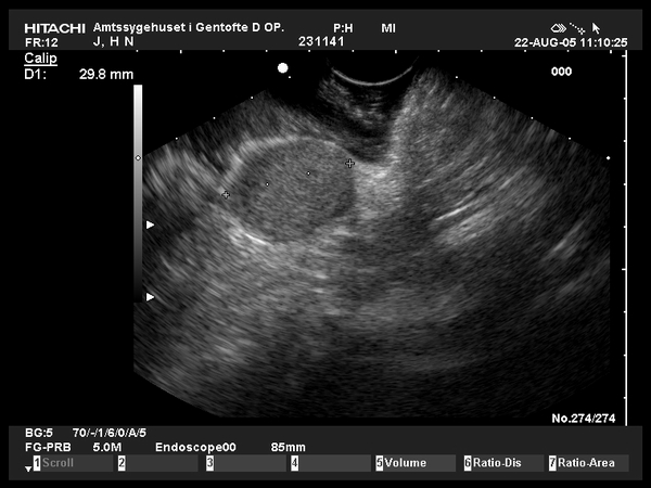
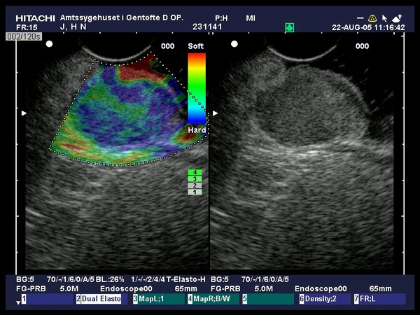
| Patient with a 3 cm gastric stromal tumor (GIST) visualized by EUS as hypoechoic, homogenous tumor, localized in the 3rd EUS layer (submucosa). Color Doppler EUS depicted discrete vascular signals in the periphery of the tumor. Elastography showed a relatively hard, in-homogenous tumor. Due to the insufficient visualization of the muscularis propria (4th EUS layer) and due to the indeterminate malignant potential, the patient was submitted for surgical resection (provide by Prof. Dr. Peter Vilmann, Copenhagen). |
 |
 |
| |
More images / movies:
|
|
|
|


