| Advanced esophageal carcinoma |
Sep 9, 2005 |
|
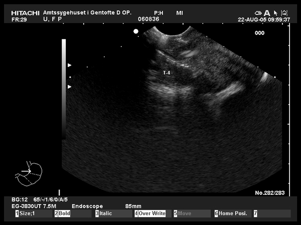
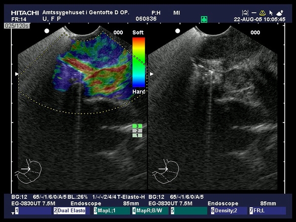
| Advanced esophageal squamous carcinoma at 36 cm distance from the incisors, staged by EUS as T4N1M1. The tumor was hypoechoic invading all layers of the esophageal wall, including the nearby pleura. A 20 mm lymph node node was visualized by EUS near the left carotid artery, confirmed by EUS-FNA as distant metastasis. Elastography guided by EUS showed the esophageal tumor as moderate hard (blue green pattern) as compared to surrounding tissues (provided by Prof. Dr. Peter Vilmann, Copenhagen). |
 |
 |
| |
More images / movies:


|
|
|
| Advanced esophageal carcinoma |
Sep 9, 2005 |
|
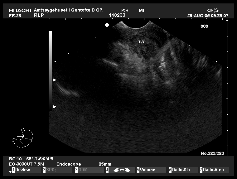
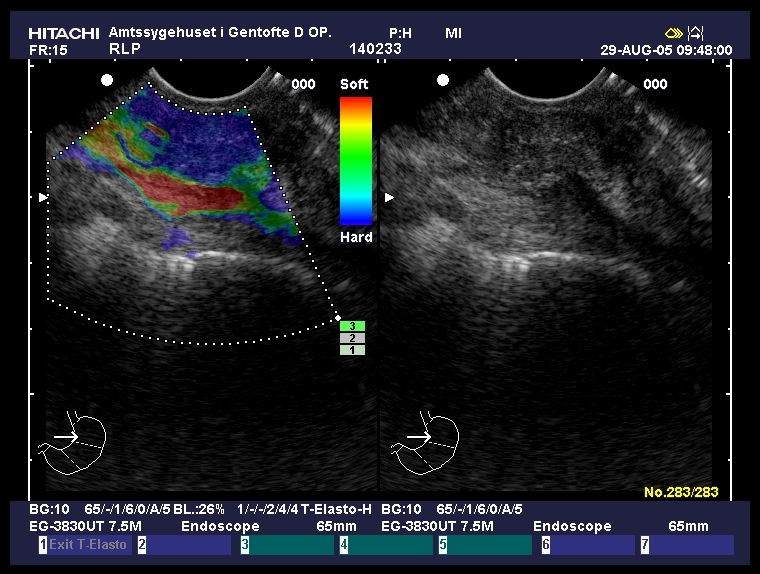
| Advanced esophageal squamous carcinoma at 34 cm distance from the incisors, staged by EUS as T3N1M1. The tumor was hypoechoic invading all layers of the esophageal wall, including the serosa. A suspicious lymph node node was visualized by EUS in the cervical area at 20 cm distance from the incisors, confirmed by EUS-FNA as distant metastasis. Elastography guided by EUS showed a uniform hard tumor (provided by Prof. Dr. Peter Vilmann, Copenhagen). |
 |
 |
| |
More images / movies:
|
|
|
|


