| Serous cystadenoma |
Feb 11, 2009 |
|
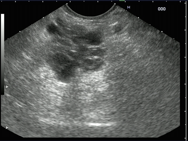
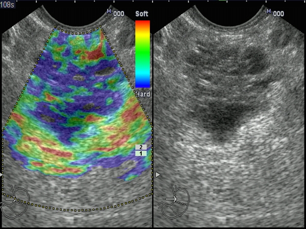
| A 71-years-old male patient, diagnosed by CT and abdominal ultrasonography with a pancreatic multicystic mass. EUS elastography revealed a mixed hardness (predominantly blue-green) of the multilocular cystic lesion, indicating a hard mass, despite the cystic structure. |
 |
 |
| |
More images / movies:

|
|
|
| Pancreatic tail cystadenocarcinoma |
Sep 9, 2005 |
|
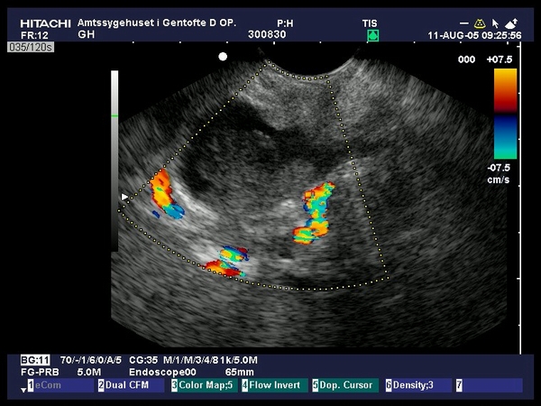
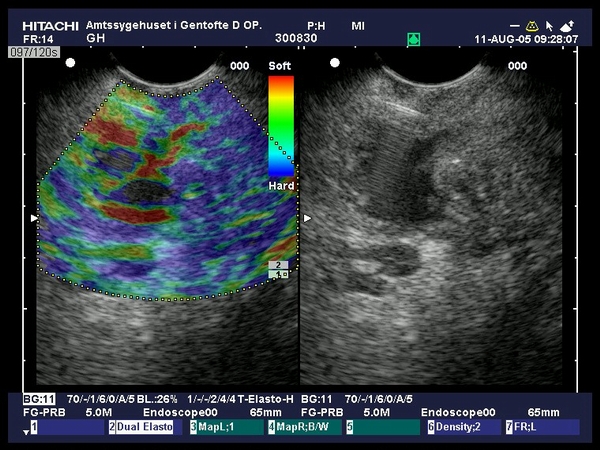
| Large cystic pancreatic tumor visualized by EUS at the level of the pancreatic tail. Color Doppler EUS showed the hypoechoic tumor embedding the splenic artery,while the splenic vein was not visualized. Elastography showed a hard tumor, as compared to the neighboring soft tissues. The diagnosis was confirmed by EUS-guided aspiration of the cystic part (high CEA values), followed by EUS-guided FNA from the solid part, which confirmed the diagnosis of malignancy (provided by Prof. Dr. Peter Vilmann, Copenhagen). |
 |
 |
| |
More images / movies:

|
|
|
|


