| Early pancreatic adenocarcinoma |
Feb 23, 2009 |
|
| Small pancreatic adenocarcinoma (20 mm) confirmed by EUS-guided FNA and visualized by EUS elastography as a hard mass (consistently blue) invading the intrapancreatic portion of the common bile duct. |
 |
 |
| |
More images / movies:

|
|
|
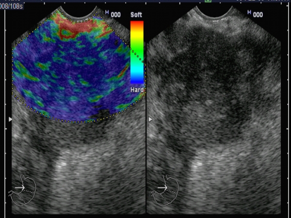
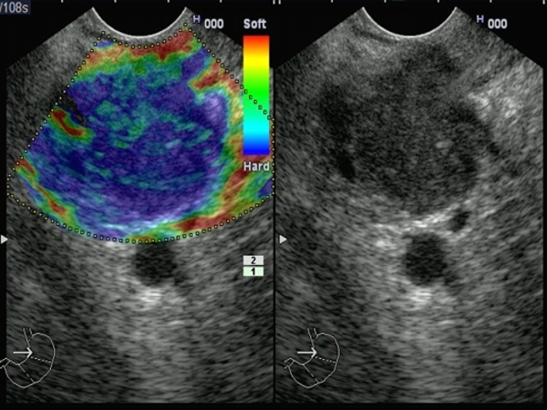
| Pancreatic body adenocarcinoma |
Feb 22, 2009 |
|
Large pancreatic body mass, in a 72-years-old man admitted with epigastric pain, weight loss and recent onset diabetes.
The diagnosis of pancreatic adenocarcinoma was confirmed by EUS-guided FNA. EUS elastography showed a hard mass (predominantly blue). |
 |
 |
| |
More images / movies:
|
|
|
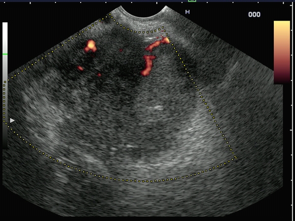
| Pancreatic adenocarcinoma |
Feb 22, 2009 |
|
| Hard (predominantly blue-green) pancreatic head mass, visualized by EUS elastography in a woman presenting with progressive jaundice. The mass was invading completely the pancreatico-duodenal artery, the distal portion of the common bile duct and pancreatic duct. The diagnosis of pancreatic adenocarcinoma was confirmed by EUS-guided FNA. |
 |
 |
| |
More images / movies:
|
|
|
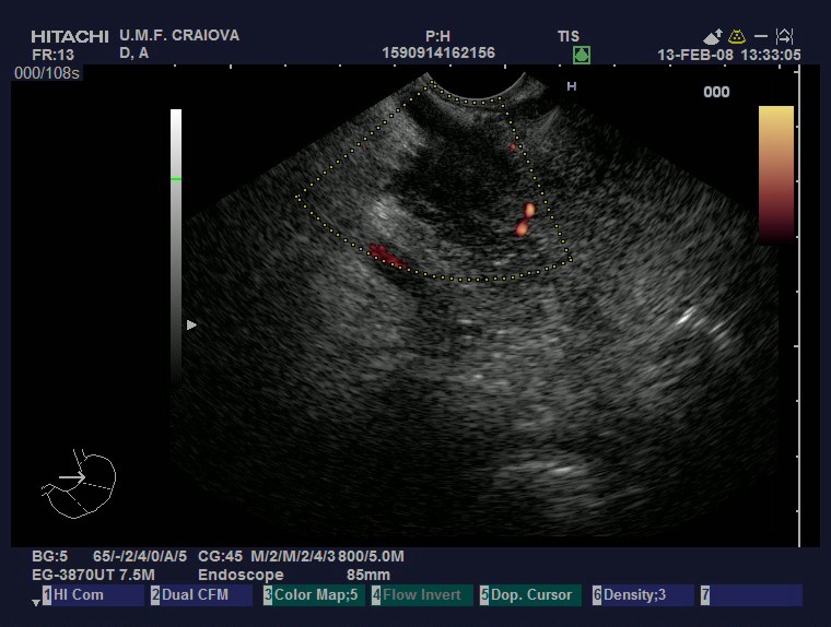
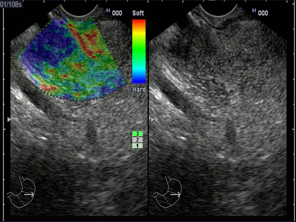
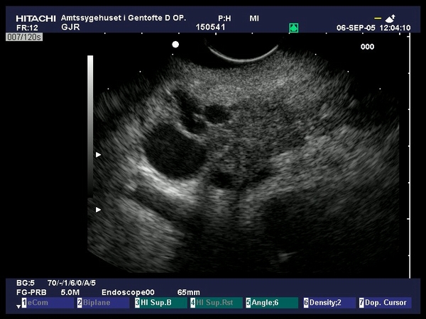
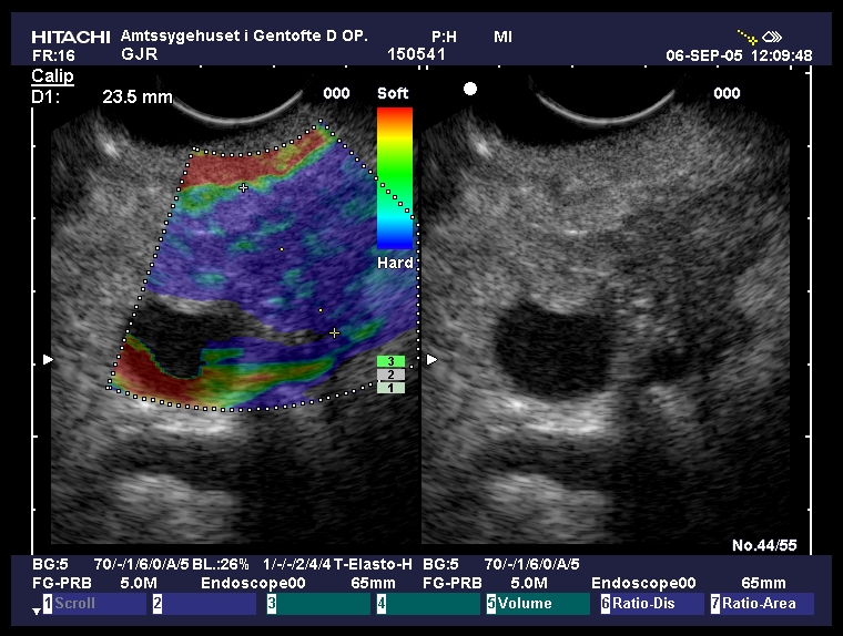
| Pancreatic head adenocarcinoma |
Sep 10, 2005 |
|
| Pancreatic head tumor mass visualized by EUS as a 3 cm hypoechoic mass at the level of the pancreatic head, with dilatation of the common bile duct and posterior invasion of the portal vein. Elastography showed a hard mass (homogenous blue pattern) as compared with the surrounding soft tissues. The diagnosis was confirmed by EUS-guided FNA with multiple passes (Provided by Prof. Dr. Peter Vilmann, Dr. Adrian Saftoiu). |
 |
 |
| |
More images / movies:

|
|
|
|


