| Pseudotumoral chronic pancreatitis |
Feb 24, 2009 |
|
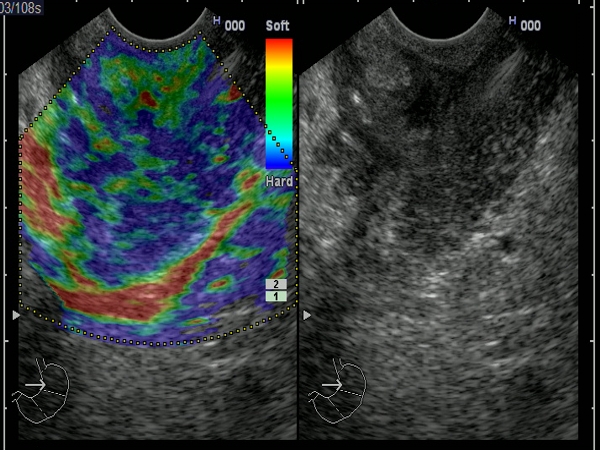
| Pseudotumoral pancreatic head mass visualized by EUS elastography with a mixed elasticity (blue-green pattern). EUS-guided FNA showed only benign canalicular cells. Long-term follow-up of more than 12 months confirmed the benign nature and absence of malignancy. |
 |
 |
| |
More images / movies:

|
|
|
| Chronic pancreatitis |
Feb 23, 2009 |
|
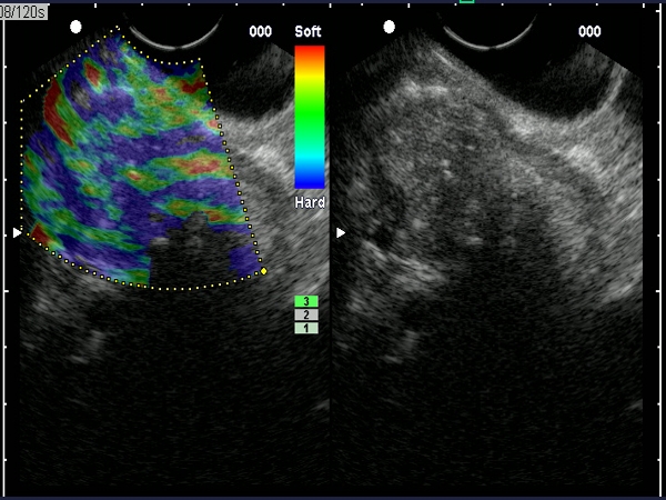
| Chronic calcifying pancreatitis visualized during EUS elastography with a mixed pattern (blue-green), indicating a mixed hardness or compressibility. |
 |
 |
| |
More images / movies:
|
|
|
| Chronic pancreatitis |
Feb 23, 2009 |
|
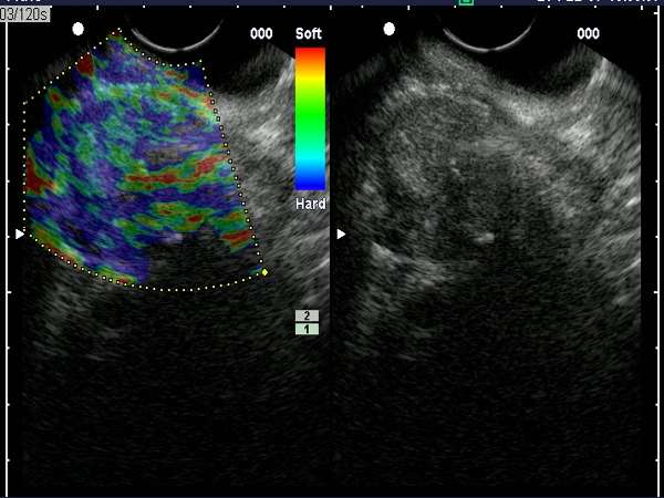
| Chronic calcifying pancreatitis visualized during EUS elastography with a mixt pattern indicating a mixed compressibility and hardness. |
 |
 |
| |
More images / movies:
|
|
|
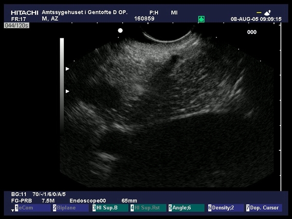
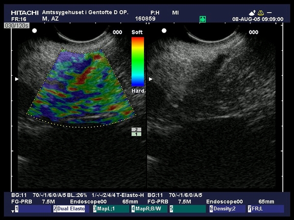
| Chronic pancreatitis |
Sep 9, 2005 |
|
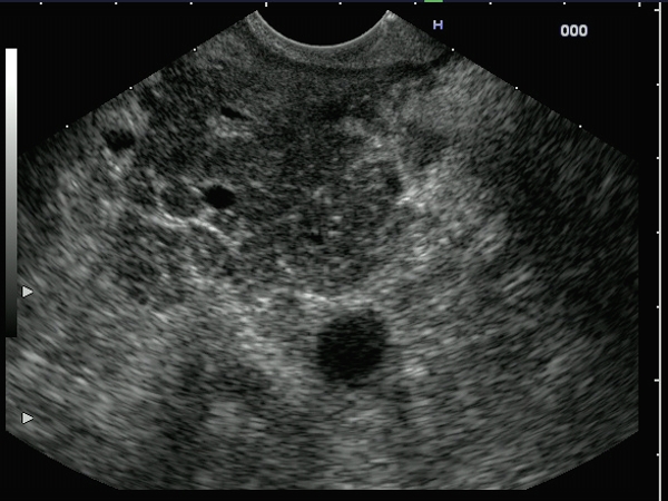
| Patient with chronic upper abdominal pain examined by EUS for a suspicion of common bile duct stones. EUS visualized an in-homogenous pancreas, with hyperechoic strands and foci, with a visible Wirsung with hyperechoic margins (criteria of early chronic pancreatitis). Elastography showed a moderate hard in-homogenous pattern (scattered blue-green signals) over the pancreatic area (Provided by Prof. Dr. Peter Vilmann, Copenhagen). |
 |
 |
| |
More images / movies:

|
|
|
|


