| |
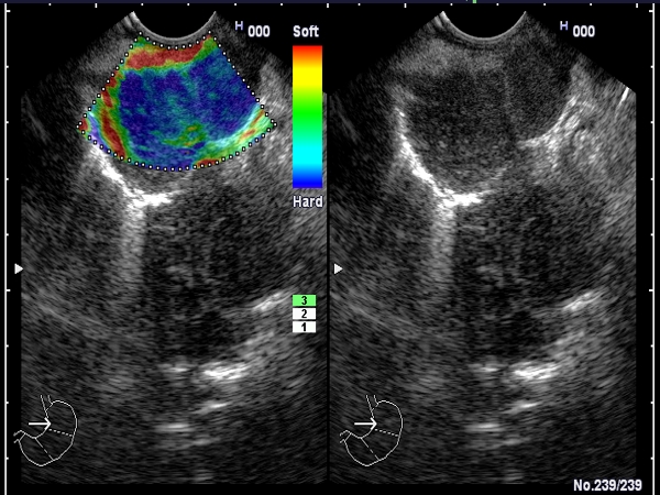
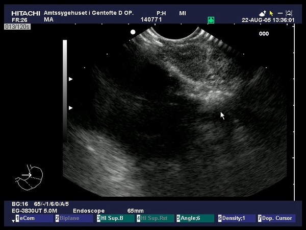
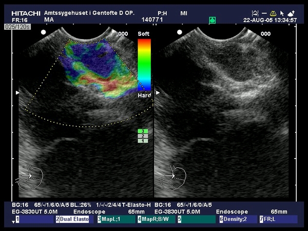
| Multiple mediastinal lymph nodes, depicted as hard by the EUS elastography software. The diagnosis of malignancy was confirmed by EUS-guided FNA from the lymph nodes located in the aortopulmonary and subcarinal space. |
 |
 |
| |
|
|
|
| Advanced esophageal carcinoma |
Sep 9, 2005 |
|
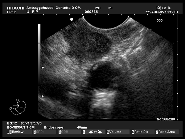
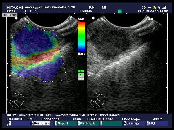
| Patient with biopsy-proven squamous cell esophageal carcinoma of the lower esophagus, staged by EUS. The tumor was hypoechoic invading all normal EUS layers of the esophagus, including the pleura (T4), with nearby round, hypoechoic lymph nodes (N1). A large lymph node (20 mm) was identified near the left carotid artery, hypoechoic, in-homogenous, confirmed by EUS-guided FNA as a distant metastasis (M1). Elastography showed a moderate hard lymph node (green blue pattern) as compared to the surrounding tissues (Provided by Prof. Dr. Peter Vilmann, Copenhagen). |
 |
 |
| |
More images / movies:


|
|
|
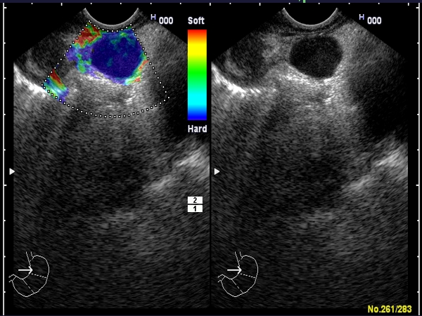
| Gastric adenocarcinoma stage IV |
Sep 9, 2005 |
|
| Young patient (34 years old) with biopsy proven gastric adenocarcinoma. EUS showed a mediastinal lymph node of 20 mm, oval, hypoechoic, in-homogenous, with irregular margins. Elastography showed an in-homogenous hard pattern (blue), suggestive for the diagnosis of malignancy. EUS-FNA was performed (2 passes) and showed atypical cells suggestive of carcinoma, confirming the diagnosis of M1 disease (provided by Prof. Dr. Peter Vilmann, Copenhagen). |
 |
 |
| |
More images / movies:
|
|
|
|


