| Mediastinal pancreatic pseudocyst |
Dec 21, 2008 |
|
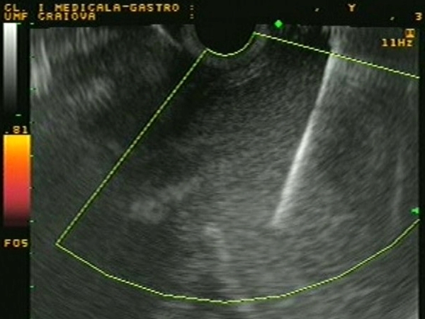
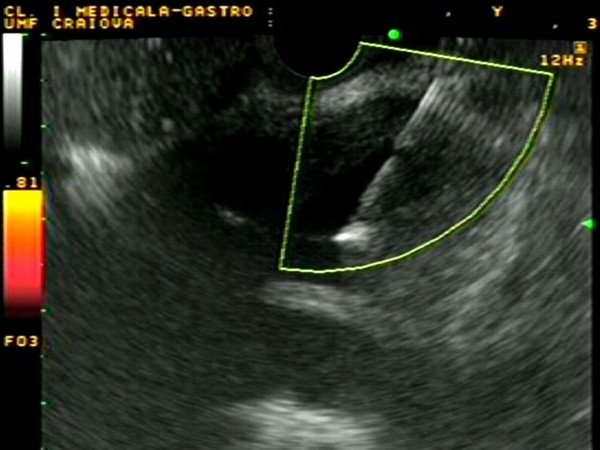
| A 30-year-old male, with known heavy alcohol abuse, was admitted in the Emergency Department complaining of intense epigastric pain, nausea and vomiting. A CT scan showed a large 15-cm pancreatic body pseudocyst that extended upwards through the diaphragmatic hiatus into the posterior mediastinum, in close connection with the descending aorta. EUS-guided drainage of the mediastinal pancreatic pseudocyst was performed through the terminal esophagus using a large-channel linear EUS scope and a one-step drainage system consisting of a diathermic catheter-guidewire assembly and a mounted 5 cm, 10 Fr stent (Giovannini Needle-wire). |
 |
 |
| |
More images / movies:


|
|
|
| Pancreatic body pseudocyst |
Aug 27, 2005 |
|
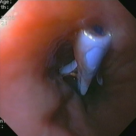
| Large pancreatic body pseudocyst of approximately 15 cm, which was compressing the stomach. The pseudocyst was drained with a Giovannini needle-wire from the stomach. After the transgastric puncture of the pseudocyst wall, a 10 Fr. stent was inserted over the needle-wire, with good drainage of the pseudocyst fluid inside the stomach. The stent was removed after 6 weeks with no complications. |
 |
 |
| |
More images / movies:
|
|
|
| Pancreatic head pseudocyst |
Feb 13, 2005 |
|
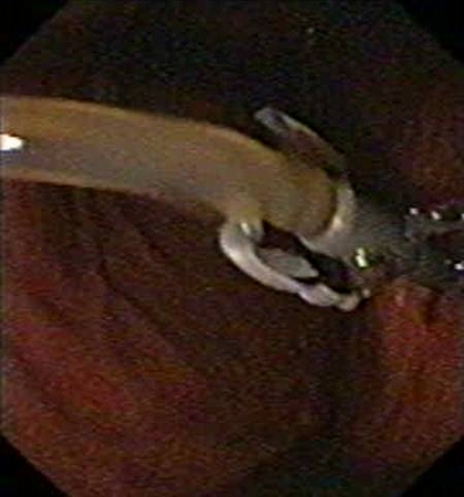
| Patient with a pancreatic head pseudocyst punctured from the duodenum with a dedicated needle-wire, covered by a catether. Contrast injection was monitored by power Doppler EUS. A 10 Fr stent was subsequently pushed over the catether. The stent can be seen on the corresponding endoscopy image. |
 |
 |
| |
More images / movies:
|
|
|
| Pancreatic head pseudocyst |
Jan 31, 2005 |
|
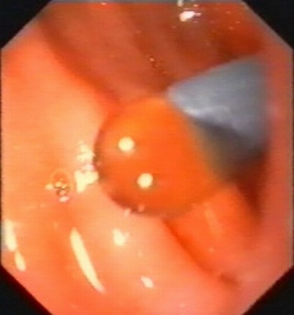
| Patient with a pancreatic head pseudocyst developed after an episode of acute biliary pancreatitis. The pseudocyst was compressing the duodenum, the common bile duct and the portal vein, causing pain and jaundice. Consequently, it was treated by EUS-guided aspiration of the content. |
 |
 |
| |
More images / movies:
|
|
|
|


