| Pancreatic head carcinoma |
Aug 30, 2005 |
|
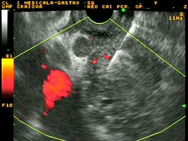
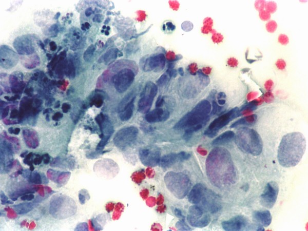
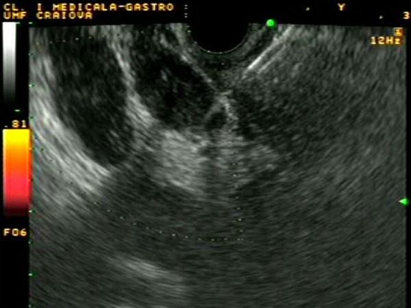
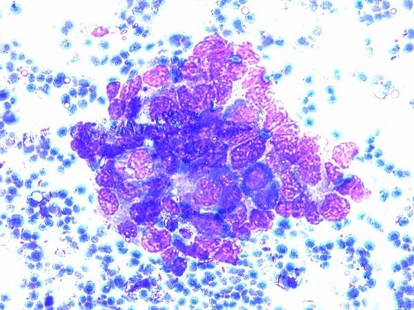
| Pancreatic head tumor mass of approximately 30 mm diameter, with collateral circulation visualized in power Doppler mode. A small lymph node (5 mm) was visible near the tumor mass. Portal vein was also evident posterior to the pancreatic head. Diagnosis was confirmed by EUS-guided FNA biopsy; the cytology smears showed clumps of atypical cells, with hypertrophic and pleomorphic nuclei, with a few erythrocytes in the background (Papanicolau smear). |
 |
 |
| |
More images / movies:

|
|
|
| Pancreatic body carcinoma |
Feb 5, 2005 |
|
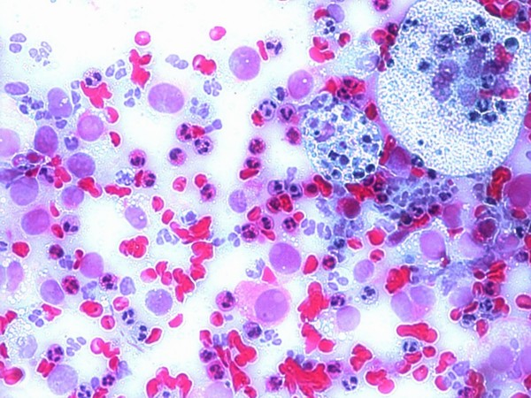
| Large pancreatic body tumor, of approximately 5 cm diameter, visualized with the ultrasound endoscope positioned in the gastric body. Diagnosis was confirmed by EUS-guided FNA biopsy; the cytology smears showed disseminated atypical cells, in a background of erythrocytes, granulocytes and macrophages (upper right).Subsequently, celiac plexus neurolysis was performed. |
 |
 |
| |
More images / movies:
|
|
|
| Advanced hilar cholangiocarcinoma |
Feb 5, 2005 |
|
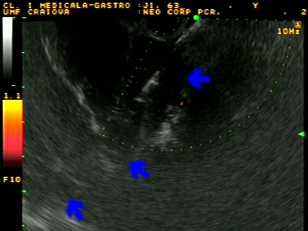
| Proximal cholangiocarcinoma (Klatskin tumor) visualized on EUS as a hypoechoic tumor mass inside the common bile duct, with complete invasion of the portal vein. The diagnosis was confirmed by EUS-guided FNA, which showed a clump of atypical cells in a background of erythrocytes. |
 |
 |
| |
More images / movies:
|
|
|
|


