| Normal azygos vein flow |
Feb 26, 2009 |
|
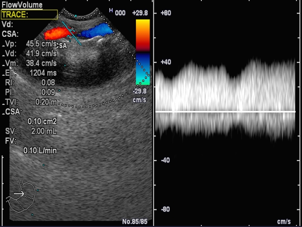
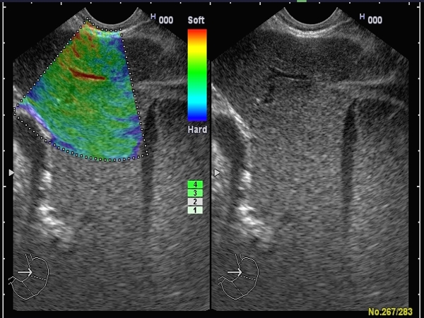
| Normal azygos vein flow (0.1 L/min) measured by pulsed Doppler EUS, from the esophagus. Normal diameter of the azygos vein, which is easily compressed with the EUS transducer. The corresponding EUS elastography image of the liver is attached (visualized from the stomach), showing a soft (normal) liver hardness. |
 |
 |
| |
|
|
|
| Increased azygos vein flow |
Feb 26, 2009 |
|
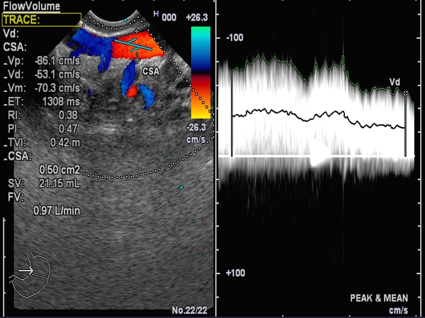
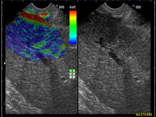
| Increased azygos vein flow (0.97 L/min) measured by pulsed Doppler EUS, from the esophagus. Increased diameter of the azygos vein, with lost compressibility induced by the EUS transducer. The corresponding EUS elastography image of the liver is attached (visualized from the stomach), showing a firm (hard) cirrhotic liver. |
 |
 |
| |
|
|
|
| Portal hypertension |
Jan 31, 2005 |
|
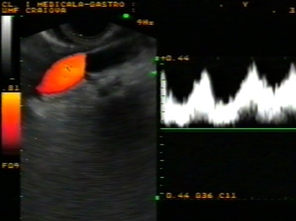
| Patient with liver cirrhosis, with increased azygos vein flow demonstrated by linear EUS in triplex mode (grey scale combined with power Doppler and pulsed Doppler ), immediately above the gastroesophageal junction. |
 |
 |
| |
|
|
|
| Portal hypertension |
Jan 31, 2005 |
|
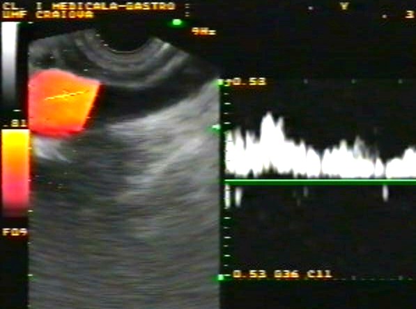
| Another patient with liver cirrhosis, with increased azygos vein flow demonstrated by linear EUS in triplex mode (grey scale combined with power Doppler and pulsed Doppler), immediately above the gastroesophageal junction. The three-layered esophageal wall is clearly visible. |
 |
 |
| |
|
|
|
|


