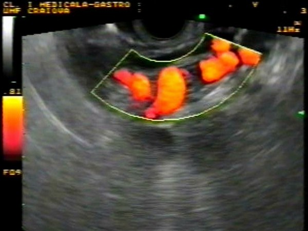
| Periesophageal collaterals |
Feb 25, 2009 |
|
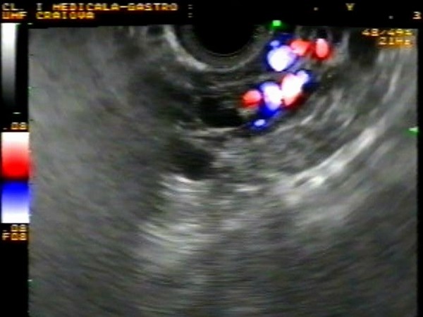
| Periesophageal collaterals visualized by EUS in color Doppler and power Doppler in a patient with known liver cirrhosis. |
 |
 |
| |
More images / movies:

|
|
|
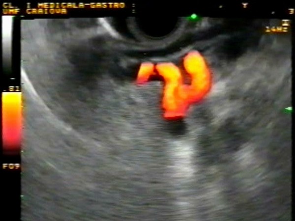
| Esophageal varices |
Jan 31, 2005 |
|
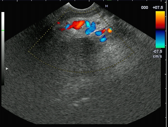
| Patient with alcoholic liver cirrhosis, with large esophageal varices demonstrated by endoscopy after an episode of upper gastrointestinal bleeding. Linear EUS clearly showed the varices in color Doppler mode, immediately above the gastroesophageal junction. |
 |
 |
| |
|
|
|
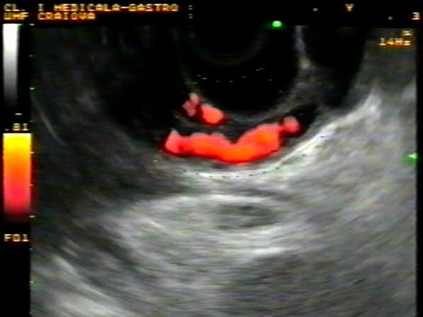
| Paraesophageal collaterals |
Jan 31, 2005 |
|
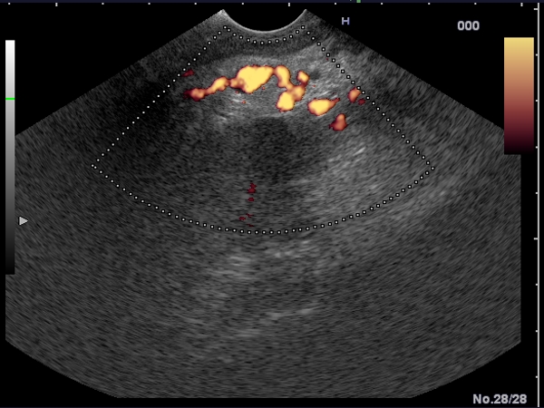
| Patient with alcoholic liver cirrhosis, with large esophageal varices eradicated after 4 sessions of endoscopic band ligation after an episode of upper gastrointestinal bleeding. Linear EUS clearly showed the paraesophageal collaterals in power Doppler mode, external of the muscularis propria of the esophagus. |
 |
 |
| |
|
|
|
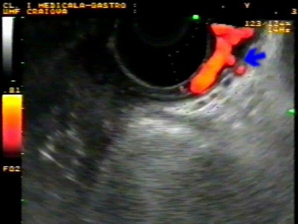
| Perforant veins |
Jan 31, 2005 |
|
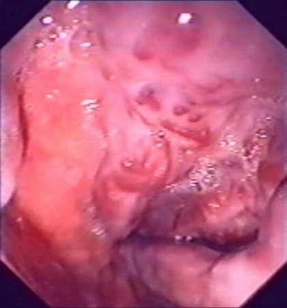
| Patient with viral liver cirrhosis, with large esophageal varices which persisted after several sessions of endoscopic band ligation, performed in an interval of 6 months. Linear EUS showed the perforant veins (blue arrow) in power Doppler mode, at 35 cm from the incisors. Both esophageal varices and paraesophageal collaterals were visualized in power Doppler EUS. |
 |
 |
| |
|
|
|
|


