| Liver metastases in pancreatic cancer |
Feb 15, 2009 |
|
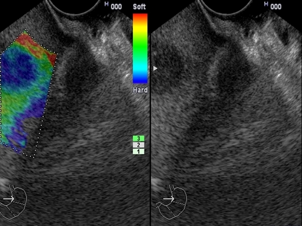
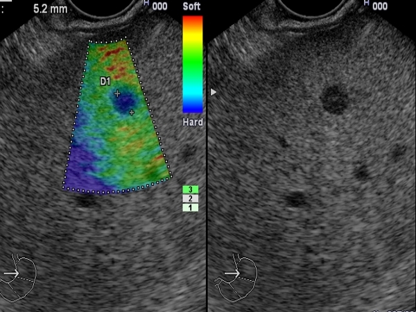
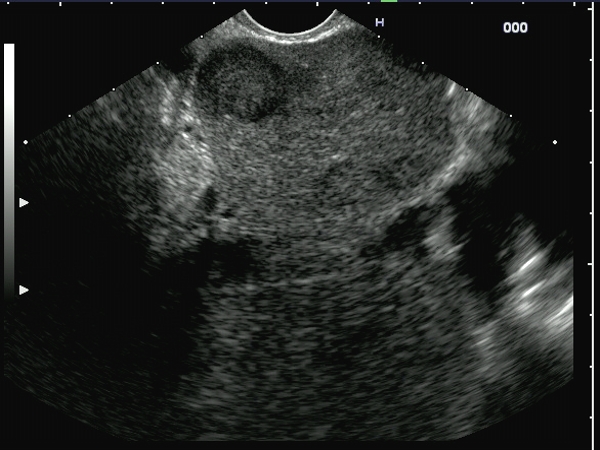
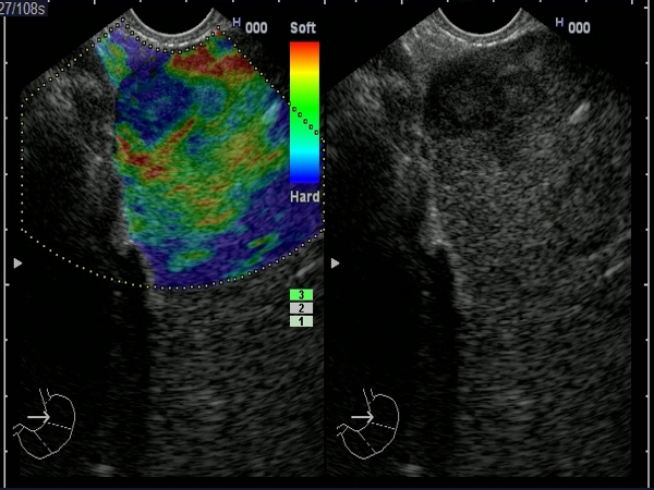
| A patient with pancreatic cancer examined by EUS for diagnosis and staging. One hypoechoic lesion without power Doppler signal was visualized in the left liver lobe, with a hard appearance (blue) during EUS elastography, suggesting the diagnosis of liver metastases. |
 |
 |
| |
More images / movies:

|
|
|
| Liver metastases in pancreatic cancer |
Feb 15, 2009 |
|
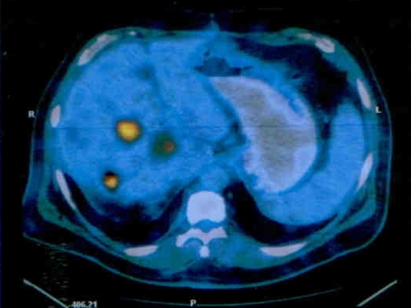
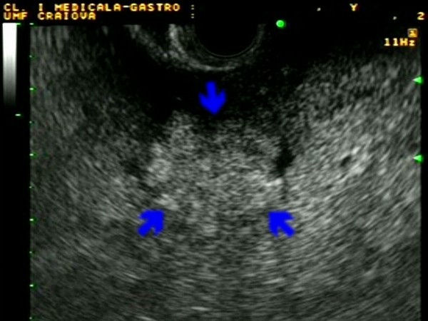
| Hard liver masses (5-10 mm in diameter) in a patient diagnosed by EUS-guided FNA with pancreatic cancer. Hybrid PET-CT examination revealed the same aspect of the liver, suggestive of multiple liver metastases. |
 |
 |
| |
More images / movies:
|
|
|
| Liver metastases in lung cancer |
Feb 15, 2009 |
|
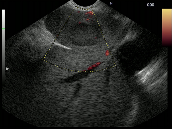
| Patient with right lung carcinoma examined by EUS for staging. Multiple hypoechoic lesions were visualized in the left liver lobe, with a hard appearance during EUS elastography. The diagnosis of malignancy was confirmed by EUS-guided FNA. |
 |
 |
| |
More images / movies:

|
|
|
| Gastric adenocarcinoma |
Jan 31, 2005 |
|
| Patient diagnosed with advanced gastric adenocarcinoma, with multiple left liver lobe metastases, visualized during EUS as hyperechoic nodules with irregular margins and peripheral halo. The left liver lobe metastases were confirmed by EUS-guided FNA biopsy. |
 |
 |
| |
More images / movies:
|
|
|
|


