| Distal cholangiocarcinoma |
Feb 10, 2009 |
|
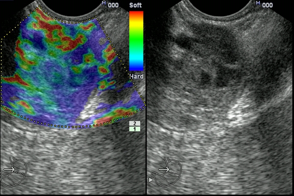
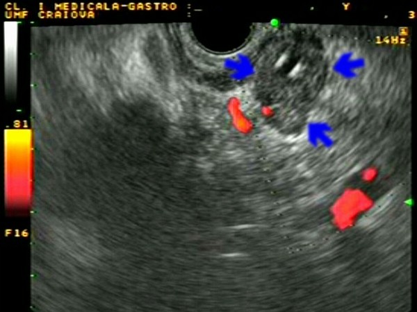
| Patient with a large hypoechoic mass (30 mm) visualized in the distal part of the common bile duct. The diagnosis o cholangiocarcinoma was confirmed by EUS-guided FNA. EUS elastography revealed the hard, but mixed appearance (predominantly blue) of the tumor. |
 |
 |
| |
More images / movies:

|
|
|
| Distal cholangiocarcinoma |
Aug 27, 2005 |
|
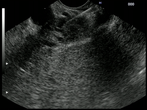
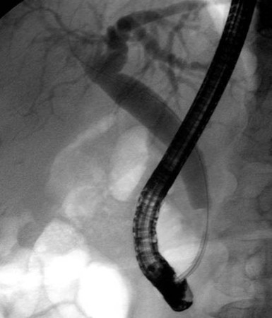
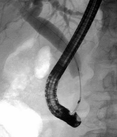
| Small hypoechoic mass (10/8 mm) visualized in the terminal part of the common bile duct, with thickening of the walls and loss of the normal wall structure. As brush cytology during a previous ERCP was negative for malignancy, the diagnosis was confirmed by EUS-guided FNA. ERCP showed a tight stricture in the terminal part of the CBD, with subsequent dilatation of intra and extrahepatic ducts. A 12 Fr. stent was consequently inserted. |
 |
 |
| |
More images / movies:
|
|
|
| Small distal cholangiocarcinoma |
Feb 8, 2005 |
|
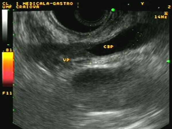
| Small distal cholangiocarcinoma visualized on EUS as a hypoechoic tumor, in the intrapancreatic terminal part of the CBD, surrounding the stent inserted during previous ERCP. EUS-FNA was negative for atypical cells; however, brush cytology from the bile duct was positive, and the patient was referred for radical duodeno-pancreatectomy. |
 |
 |
| |
More images / movies:

|
|
|
|


