| 3D advanced pancreatic adenocarcinoma |
Feb 22, 2009 |
|
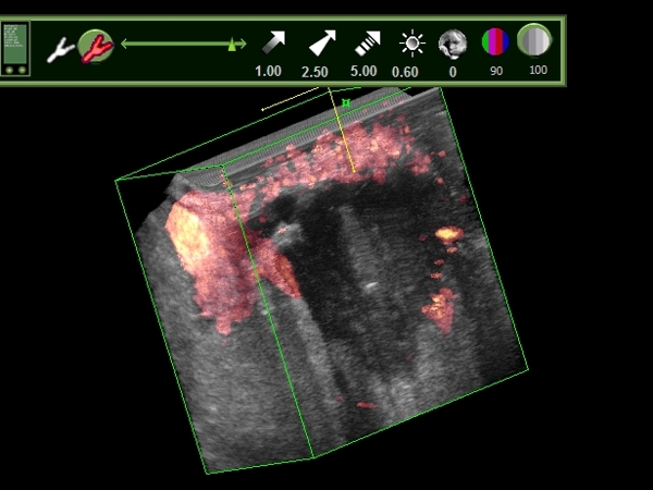
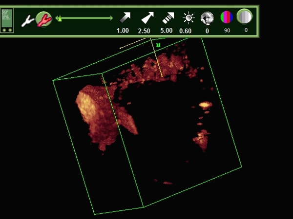
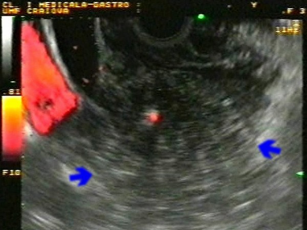
| Large pancreatic body adenocarcioma confirmed by EUS-guided FNA, with important collateral circulation visualized in power Doppler mode after contrast-enhancement (Sonovue). A tridimensional (3D) reconstruction eased the visualization of the relationship between the tumor and splenomesenteric confluent. |
 |
 |
| |
More images / movies:

|
|
|
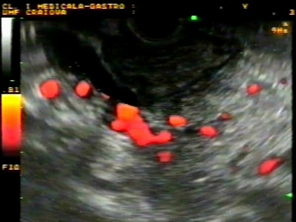
| Early pancreatic adenocarcinoma |
Feb 22, 2009 |
|
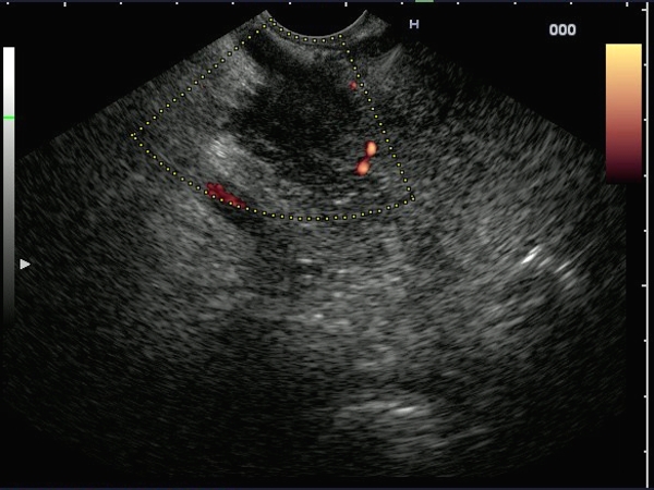
| Small hypoechoic mass (20 mm), invading the common bile duct in a patient with jaundice with recent onset. The mass had absent power Doppler signals inside, and was located nearby the splenomesenteric confluence, without invading it. The diagnosis of adenocarcinoma was confirmed by EUS-guided FNA. |
 |
 |
| |
More images / movies:
|
|
|
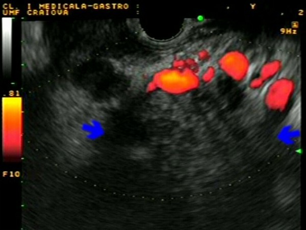
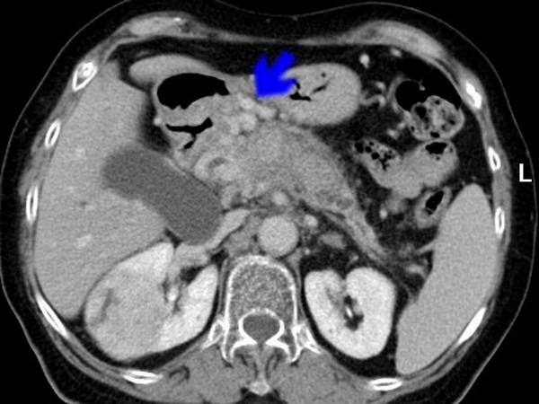
| Peripancreatic collaterals |
Aug 26, 2005 |
|
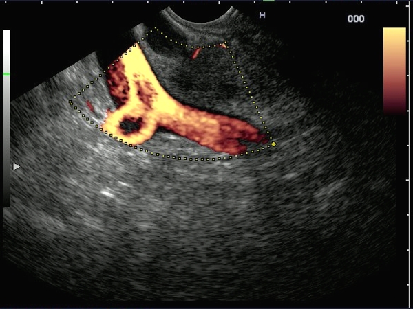
| Large collaterals visualized by power Doppler EUS, surrounding a large hypovascular pancreatic body tumor. The collaterals were also visualized on the CT scan, anterior to the pancreatic tumor. Despite the presence of collaterals, the diagnosis was confirmed by EUS-FNA. |
 |
 |
| |
More images / movies:

|
|
|
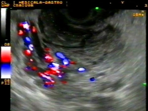
| Cavernomatous transformation of the portal vein |
Jan 25, 2005 |
|
| Collaterals surrounding the terminal part of the common bile duct, visualized by conventional color and power Doppler EUS, with the ultrasound scope positioned in the duodenal bulb. The normal portal vein was not visualized. |
 |
 |
| |
|
|
|
| Invasion of the portal vein |
Jan 25, 2005 |
|
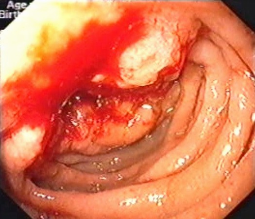
| Large pancreatic head tumor mass of approximately 50 / 45 mm, with invasion of the terminal common bile duct and complete invasion of the portal vein. Duodenal invasion at the level of papilla major was also demonstrated by upper GI endoscopy; diagnosis of adenocarcinoma was confirmed by biopsies. |
 |
 |
| |
|
|
|
|


