| Pancreatic adenocarcinoma |
Feb 22, 2009 |
|
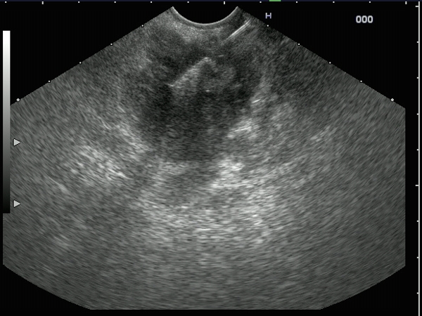
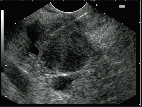
| Hypoechoic, inhomogenous tumor mass, located at the level of the pancreatic head in a 70-years-old woman with progressive jaundice. The diagnosis of pancreatic adenocarcinoma was confirmed by EUS-guided FNA. Both cytology and cell-blocks with immunohistochemistry (AE1 / AE3) were consistent with the diagnosis of pancreatic adenocarcinoma. |
 |
 |
| |
More images / movies:


|
|
|
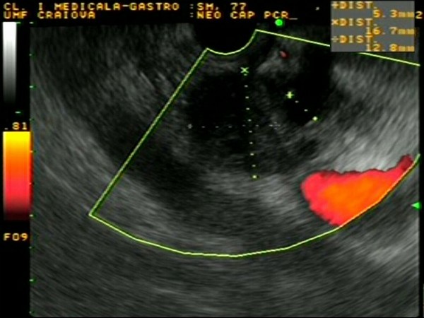
| Advanced pancreatic body adenocarcinoma |
Feb 22, 2009 |
|
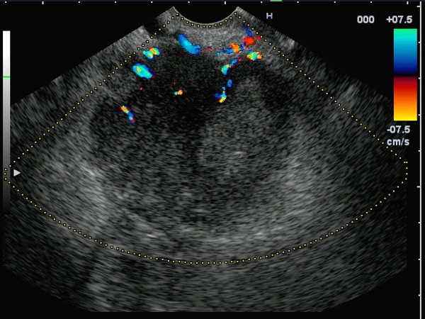
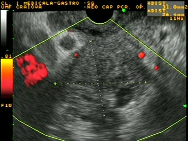
| Large pancreatic body mass, in a 72-years-old man admitted with epigastric pain, weight loss and recent onset diabetes. Collateral circulation was visualized in color Doppler and power Doppler. The diagnosis of pancreatic adenocarcinoma was confirmed by EUS-guided FNA. Cell blocks examination showed a poorly differentiated adenocarcinoma, while immunohistochemistry was positive for CA 19-9. |
 |
 |
| |
More images / movies:
|
|
|
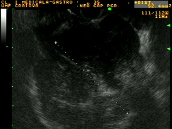
| Pancreatic adenocarcinoma |
Feb 15, 2009 |
|
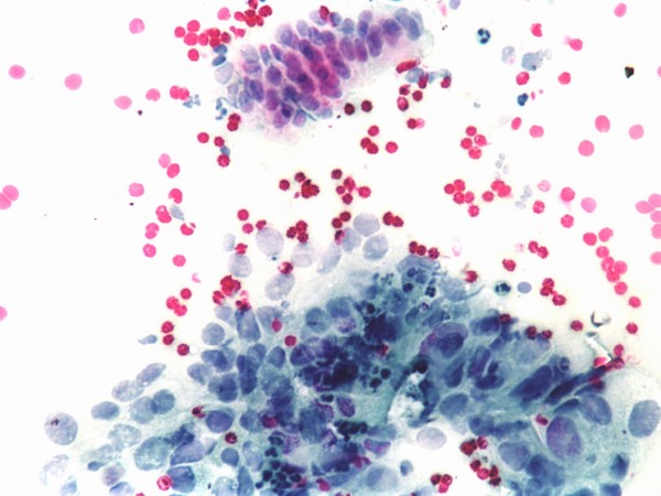
| Hypoechoic pancreatic head mass of approximately 40/30 mm, visualized by EUS in a 70-years-old women, presenting with progressive jaundice. The mass was invading completely the distal portion of the common bile duct and pancreatic duct, as well as the pancreaticoduodenal artery. The diagnosis was confirmed by EUS-guided FNA, showing clumps of atypical cells. |
 |
 |
| |
More images / movies:
|
|
|
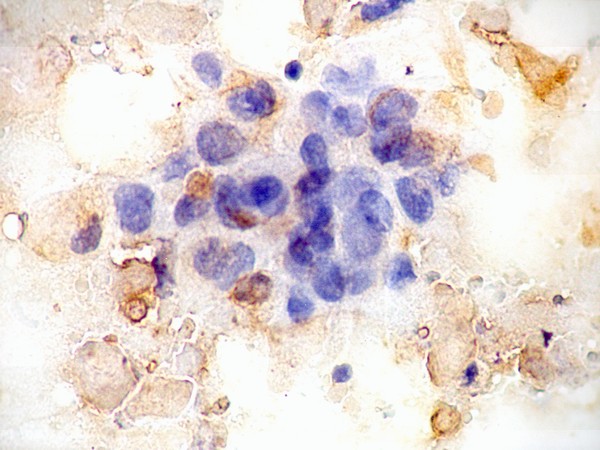
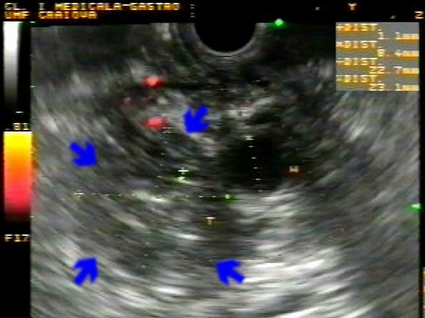
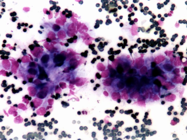
| Pancreatic head carcinoma |
Aug 30, 2005 |
|
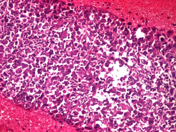
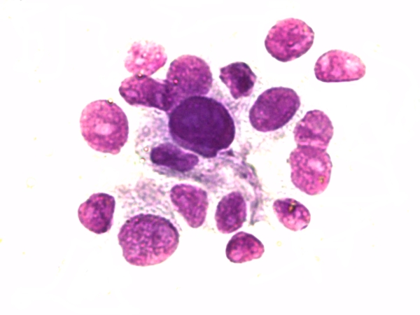
| Pancreatic head tumor mass of approximately 30 / 25 mm, with collateral circulation visualized in power Doppler mode. The mass was invading the portal vein and the terminal part of the common bile duct. Diagnosis was confirmed by EUS-guided FNA biopsy. The cytology smears showed clumps of atypical cells (bottom), with a small island of normal acinar cells (top), in a background of erythrocytes. |
 |
 |
| |
More images / movies:
|
|
|
| Small pancreatic head carcinoma |
Mar 2, 2005 |
|
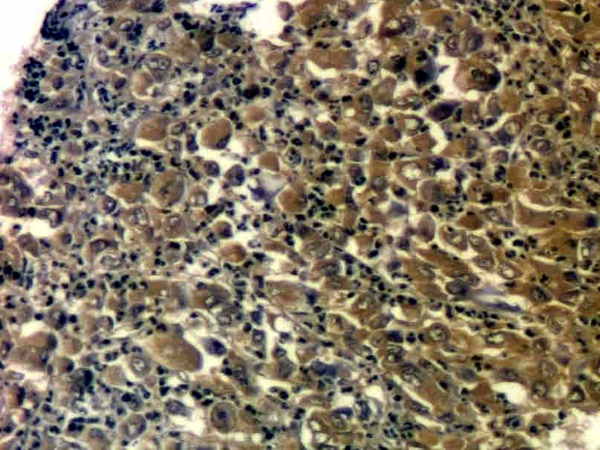
| Hypoechoic tumor mass of approximately 20 / 15 mm at the level of the pancreatic head, with retrograde dilatation of the main pancreatic duct. Diagnosis was confirmed by EUS-guided FNA biopsy. The cytology smears showed clumps of atypical cells, while immunocytochemistry was positive for cytokeratins AE1/AE3 inside the tumoral cells. The patient was subsequently referred for radical duodeno-pancreatectomy. |
 |
 |
| |
More images / movies:
|
|
|
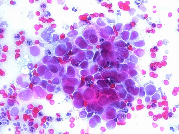
| Pancreatic body carcinoma |
Feb 5, 2005 |
|
| Large pancreatic body tumor, of approximately 5 cm diameter, visualized with the ultrasound endoscope positioned in the gastric body. Diagnosis was confirmed by EUS-guided FNA biopsy; the cytology smears showed clumps of atypical cells. Furthermore, celiac plexus neurolysis was performed. |
 |
 |
| |
More images / movies:
|
|
|
| Pancreatic head carcinoma in chronic pancreatitis |
Jan 31, 2005 |
|
| Patient with known chronic pancreatitis with inhomogenous pancreatic head with calcifications. A suspicion of a pancreatic head tumor mass was raised based on CT findings. This was also suggested by the absence of power Doppler signals inside the tumor mass. Diagnosis was confirmed by EUS-guided FNA biopsy. The smears showed clumps of atypical cells in a background of erythrocytes. |
 |
 |
| |
|
|
|


